


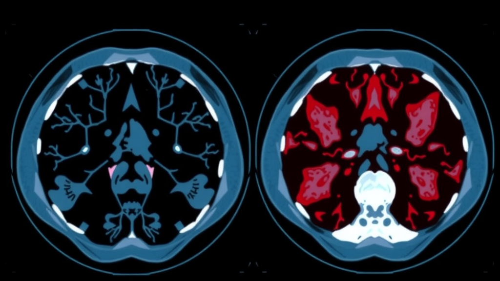
Deep in the retroperitoneum, the crowded space at the back of the abdomen where the kidneys, pancreas, and great vessels reside, two very different cancers can grow to enormous sizes before anyone notices. Dedifferentiated liposarcoma and leiomyosarcoma are the two most common non-fatty malignant tumors of this region, and telling them apart before surgery is one of the most stubborn challenges in abdominal imaging. Both appear as large, heterogeneous soft-tissue masses on computed tomography, both affect similar patient populations, and both demand radically different surgical and oncological strategies. A new study published in BMC Medical Imaging suggests that the answer may lie not in what radiologists can see with their eyes, but in the statistical fingerprints of tumor texture that only machine learning can reliably extract.
Researchers led by Enlong Zhang and Yuan Li of Peking University Third Hospital, together with colleagues at Tsinghua University Hospital and Peking University International Hospital, developed a tumor habitat-based radiomics approach to distinguish retroperitoneal non-fatty dedifferentiated liposarcoma from leiomyosarcoma. Their retrospective study included 166 patients, 102 with dedifferentiated liposarcoma and 64 with leiomyosarcoma, all of whom underwent multiphase contrast-enhanced CT. Rather than treating each tumor as a single uniform blob, the team borrowed a concept from oncology known as intratumoral heterogeneity, the recognition that a cancer is a patchwork of biologically distinct microenvironments, each with its own cellularity, vascularity, necrosis, and stromal composition.
The technical core of the method is elegantly simple. On the largest axial slice of each tumor, the researchers applied a Simple Linear Iterative Clustering algorithm, a superpixel segmentation technique originally developed for computer vision, to divide the tumor image into hundreds of small, coherent regions of similar pixel intensity. A K-means clustering step then grouped these superpixels into three habitat subregions, effectively partitioning each tumor into three zones that reflect different levels of attenuation, a radiological proxy for tissue density. The team designated these subregions ROI1, ROI2, and ROI3, with ROI2 corresponding to the intermediate-attenuation zone that often represents a transitional territory between viable cellular tissue and degenerative change.
From each habitat subregion, across three imaging phases, the non-contrast phase, the arterial phase, and the venous phase, the researchers extracted a comprehensive panel of quantitative radiomics features. These included shape descriptors such as major axis length, first-order statistics capturing the distribution of gray values, and higher-order texture features derived from gray-level co-occurrence matrices, run-length matrices, size zone matrices, dependence matrices, and neighborhood gray-tone difference matrices, as well as wavelet-transformed versions of these features that probe texture at multiple spatial scales. Feature selection was performed independently within each subregion using LASSO regression, and predictive models were then built with XGBoost, a gradient-boosted decision tree algorithm renowned for its performance on tabular biomedical data.
The results point to a clear conclusion: contrast matters. Habitat radiomics models derived from contrast-enhanced CT generally outperformed those built on non-contrast images, suggesting that the way tumor subregions take up iodinated contrast, and therefore their vascular perfusion characteristics, carries diagnostic information that plain density measurements miss. The single best-performing model was built from the venous-phase intermediate-attenuation subregion, VP-ROI2. In the testing cohort it achieved an area under the receiver operating characteristic curve of 0.834, with a 95 percent confidence interval of 0.713 to 0.950, alongside an accuracy of 0.820, a sensitivity of 0.871, and a specificity of 0.737.
The venous-phase advantage is physiologically plausible. The venous phase of a contrast-enhanced CT, typically acquired around sixty to ninety seconds after injection, captures the period of maximum enhancement in many soft-tissue tumors, when contrast has diffused into the extracellular space and reflects capillary permeability and interstitial volume. Dedifferentiated liposarcoma and leiomyosarcoma differ in their microvascular architecture and stromal composition, and those differences apparently imprint distinguishable enhancement patterns on the intermediate-attenuation habitat. Interestingly, VP-ROI2 showed only a nominally significant advantage over its non-contrast counterpart, NP-ROI2, with a P value of 0.048 that fell to an adjusted P value of 0.173 after false discovery rate correction, a statistical caution the authors appropriately acknowledge. ROI2 nevertheless emerged as the most discriminative subregion on both arterial and venous phases.
Perhaps the most compelling aspect of the study is its commitment to interpretability, a virtue not always found in machine learning medicine. Using SHAP, or SHapley Additive exPlanations, a game-theoretic framework that quantifies each feature’s contribution to individual predictions, the team identified original_shape_MajorAxisLength and wavelet-based texture features as the most influential predictors. Shape matters, which makes intuitive sense: dedifferentiated liposarcomas and leiomyosarcomas differ in their growth patterns and margins. But the prominence of wavelet texture features, which encode fine-scale spatial patterning of pixel intensities, indicates that the microscopic organization of tumor tissue, its heterogeneity at radiologically invisible scales, is what truly separates the two diagnoses. The models were built and reported following established standards, including the Image Biomarker Standardization Initiative nomenclature and the TRIPOD guidelines for prediction model reporting, and the study received an assessment of radiomics quality.
The clinical implications are significant. Today, distinguishing these sarcomas preoperatively often depends on biopsy, and even then core needle sampling of a large heterogeneous tumor can miss the diagnostic territory, a problem known as sampling error. Accurate preoperative differentiation guides the surgical plan: dedifferentiated liposarcoma demands wide clearance of the fatty component along with the dedifferentiated nodule, whereas leiomyosarcoma management hinges more on vascular involvement and multimodal planning. A noninvasive imaging biomarker that can flag the likely diagnosis before the first incision could help surgeons counsel patients, select neoadjuvant strategies, and design clinical trials that stratify patients by histological subtype rather than by the blunt label of retroperitoneal sarcoma.
The authors are careful to frame this as a promising step rather than a finished clinical tool. The cohort was retrospective and drawn from Chinese hospitals, the tumors were segmented on the largest axial slice rather than in three dimensions, and the performance figures, while respectable, leave room for improvement before the model could stand beside pathology. External validation in independent, multiethnic cohorts and prospective testing will be essential before VP-ROI2-based habitat radiomics enters routine decision-making. Still, the study offers a vivid demonstration of a broader idea now sweeping through oncological imaging: that every tumor is a landscape of habitats, and that the map of that landscape, drawn automatically from a routine contrast-enhanced CT scan, may hold answers that biopsies, radiologists, and even pathologists struggle to provide. The venous phase, long treated as a routine addendum to the arterial spectacle, turns out to be where the sarcomas reveal their secrets.
Subject of Research: Venous-phase CT-based tumor habitat radiomics for differentiating retroperitoneal non-fatty dedifferentiated liposarcoma from leiomyosarcoma
Article Title: Venous-phase CT-based tumor habitat radiomics for differentiating retroperitoneal non-fatty dedifferentiated liposarcoma from leiomyosarcoma
Article References: Zhang, E., Li, Y., Ma, L., Ji, D., Zhang, M., & Lang, N. (2026). Venous-phase CT-based tumor habitat radiomics for differentiating retroperitoneal non-fatty dedifferentiated liposarcoma from leiomyosarcoma. BMC Medical Imaging. https://doi.org/10.1186/s12880-026-02784-4
Image Credits: AI Generated
DOI: 10.1186/s12880-026-02784-4
Keywords: retroperitoneal sarcoma, dedifferentiated liposarcoma, leiomyosarcoma, radiomics, tumor habitat, intratumoral heterogeneity, computed tomography, machine learning, XGBoost, SHAP, cancer imaging, preoperative diagnosis
Cite Scienmag News
Ophelia Keating. (September 12, 2026). AI Reads CT Scans to Tell Two Deadly Sarcomas Apart. Scienmag. https://scienmag.com/ai-reads-ct-scans-to-tell-two-deadly-sarcomas-apart/
Ophelia Keating. "AI Reads CT Scans to Tell Two Deadly Sarcomas Apart." Scienmag, 12 September 2026, https://scienmag.com/ai-reads-ct-scans-to-tell-two-deadly-sarcomas-apart/. Accessed 12 September 2026.
Ophelia Keating. "AI Reads CT Scans to Tell Two Deadly Sarcomas Apart." Scienmag. September 12, 2026. https://scienmag.com/ai-reads-ct-scans-to-tell-two-deadly-sarcomas-apart/


