


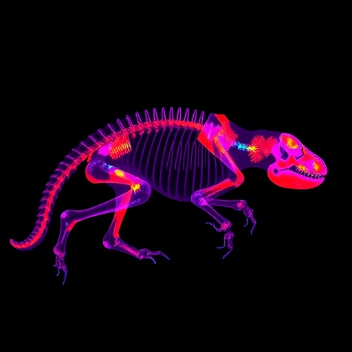
A new comprehensive review published in Environmental Chemistry Letters surveys more than three decades of X-ray fluorescence research in animal and human biological samples, charting the rise of a technique that promises non-destructive, multi-elemental quantification and imaging of potentially toxic metals and metalloids across soft tissues, hard tissues and biological fluids. Authored by Noah Casañas and Maxime Louzon of the French ecotoxicology company ENVISOL in Grenoble together with Aurélie Pelfrêne of Université de Lille, the review covers studies published between 1994 and 2026 and positions X-ray fluorescence spectrometry, widely abbreviated XRF, as an increasingly credible complement to the destructive, plasma-based techniques such as inductively coupled plasma mass spectrometry that have long dominated elemental analysis in biological matrices. The authors argue that growing industrial, agricultural and urban releases of metal(loid)s into ecosystems demand faster, less invasive and more spatially informative tools for evaluating exposure, bioaccumulation and long-term health risks, and that XRF is now mature enough to fill much of that gap, provided that standardisation and calibration challenges are decisively addressed.
The physical principle underlying XRF is deceptively simple. When a sample is irradiated with high-energy X-ray photons, inner-shell electrons in constituent atoms are ejected, and as outer electrons fall to fill the resulting vacancies, atoms emit secondary, or fluorescent, X-rays at energies and wavelengths characteristic of each element. By detecting and analysing this emitted radiation, researchers can identify and quantify elements within a sample without dissolving, digesting or otherwise destroying it. Energy-dispersive instruments resolve the fluorescent photons by energy using solid-state detectors, while wavelength-dispersive systems separate them by diffraction, achieving higher spectral resolution at the cost of greater complexity. Monochromatic excitation configurations narrow the excitation spectrum to reduce background scatter, and total reflection XRF exploits grazing-incidence optics to suppress the scattering continuum, dramatically lowering detection limits for minute liquid or thin-film specimens. Each variant carries trade-offs in sensitivity, throughput and suitability for particular biological materials, a central theme the review dissects systematically.
What has transformed the field in recent years, the authors contend, is the parallel maturation of three distinct instrumental families. Portable and handheld XRF analysers, once considered crude screening tools, now offer benchtop-grade quantification directly in the field, at the bedside, or in wildlife rehabilitation centres. Micro-focused beams deliver spatial resolution sufficient to map element distributions within tissue sections at the micrometre scale. And synchrotron sources, with their brilliance and tunability, push detection down to nanoscopic dimensions, enabling element-specific imaging of individual organelles, nanoparticle agglomerates and cellular substructures. The review documents how this technological spread has opened applications spanning environmental toxicology, medical diagnostics, veterinary science, wildlife monitoring and forensic authentication, often in contexts where conventional laboratory workflows would destroy irreplaceable specimens or simply arrive too late to inform a decision.
Among the most striking case studies assembled in the review are field deployments of portable XRF on living animals. Researchers have measured bone lead in live California condors, an endangered scavenger species chronically threatened by ingested lead ammunition fragments, allowing conservation teams to triage individuals for chelation therapy without sacrificing a single bird. Comparable in vivo measurements have been performed on Australian wedge-tailed eagles and other raptors, and on duck species examined two decades after a ban on lead shot, revealing that legacy contamination persists in food webs long after regulatory action. In humans, in vivo K-line XRF of bone lead has accrued three decades of epidemiological validation, serving as a cumulative-exposure biomarker in studies of lead poisoning in children, declining population bone lead in Canada, and occupational cohorts. The technique has even been explored clinically for monitoring iron overload in patients with thalassemia and hemochromatosis through portable skin measurements.
At the opposite end of the resolution spectrum, synchrotron-based micro- and nano-XRF has delivered biological insights that bulk chemistry cannot access. The review catalogues studies that mapped mercury selenide deposits in striped dolphin tissues, traced copper-rich aggregates in ageing rodent brain astrocytes, localised manganese in dopaminergic neurons of the substantia nigra, and revealed gadolinium retention in the inflamed brains of mice given MRI contrast agents. In ecotoxicology, synchrotron imaging has tracked cerium dioxide nanoparticles colocalising with pharyngeal deformities in the model nematode Caenorhabditis elegans, distinguished waterborne from dietary zinc accumulation in Daphnia magna, and visualised arsenic distribution in earthworm coelomic fluid as a proxy for soil bioavailability. Several studies combined XRF with X-ray absorption spectroscopy to simultaneously determine where elements sit and in what chemical form, a capability the authors highlight as essential, since toxicity depends far more on speciation than on total concentration.
Forensic science emerges as an unexpected beneficiary. Because elemental profiles of mineralised tissues vary systematically between species and even between individuals raised in different environments, handheld XRF has been used to distinguish Asian from African elephant tusks, to classify dugong tusks against other species, and to differentiate the bones of humans, elephants, dogs and dolphins for species identification. Hybrid statistical classifiers built on XRF elemental data can separate genuine ivory from counterfeit products, and elemental signatures have been proposed for tracing provenance in illegal wildlife trade seizures. These applications exploit precisely the properties that make XRF attractive in the field: speed, portability, non-destructiveness, and simultaneous multi-element readout, all without any chemical preparation that might alter evidentiary material.
The review is candid about the obstacles that still separate routine practice from these headline successes. Matrix effects, the alteration of measured fluorescence intensities by the composition of the surrounding tissue, are particularly severe in heterogeneous biological specimens, where water content, lipid fraction, mineral density and organic matrix vary dramatically between, say, fatty liver, cortical bone and blood serum. Calibration remains the persistent weak point: certified reference materials matching the matrix and concentration range of real animal tissues are scarce, and inter-laboratory comparisons reveal discrepancies that standard addition methods, fundamental-parameter corrections and cross-validation against ICP-MS are only beginning to close. Detection limits, typically adequate for major and minor elements, remain marginal for trace toxicants in soft tissues, where concentrations can fall to the low microgram-per-gram level or below. The authors also stress that XRF cannot by itself provide isotope information or chemical speciation, reinforcing its designated role as a complement rather than a replacement for mass-spectrometric and chromatographic techniques.
Sample preparation strategy, the review shows, is where analytical philosophy diverges most sharply across XRF configurations. Minimal-preparation approaches, such as direct measurement of intact bone, teeth, hooves, shells or hair, preserve specimen integrity and enable in vivo work but constrain detection limits and demand careful correction for geometry and overlying tissue. Cryosectioning and freeze-drying of soft tissues yield smooth, homogeneous surfaces amenable to micro-imaging but introduce handling steps that risk contamination or elemental redistribution. Total reflection XRF accommodates tiny droplets of serum or digested tissue, while benchtop and synchrotron microprobes require thin, flat sections mounted on clean supports. Radiation damage, generally negligible for static measurements, becomes a genuine consideration for extended synchrotron scans of living or delicate specimens, and the review cites work on plants demonstrating measurable in vivo effects that biological researchers must anticipate.
Trends across the 1994–2026 corpus reveal both consolidation and diversification. Early studies concentrated on validated in vivo bone lead measurement in humans and laboratory animals; the mid-2000s saw expansion into wildlife ecotoxicology and forensic identification; and the 2010s brought an explosion of synchrotron imaging applied to nanoparticle toxicology, neurodegeneration and developmental biology. The most recent years are marked by the migration of benchtop technology into field settings and by hybrid workflows that pair rapid XRF screening with confirmatory ICP-MS or laser ablation mass spectrometry on selected samples. Gaps persist, the authors note, in the analysis of biological fluids, in quantitative three-dimensional imaging, and in harmonised reporting of detection limits and uncertainty, all of which hamper cross-study comparison and regulatory acceptance.
The review’s concluding argument is pragmatic rather than triumphal. XRF, the authors write, offers unique opportunities for non-destructive multi-elemental analysis, environmental monitoring, medical and veterinary application and forensic authentication, but its credibility rests on improved standardisation, rigorous calibration protocols and systematic cross-validation against established methods. The authors point to green analytical chemistry principles, reduced solvent use, minimal sample destruction and the ability to move the laboratory to the sample, as a strategic advantage that aligns XRF with the broader sustainability agenda of modern analytical science. If the community can deliver the metrological groundwork, the technology stands to become an indispensable first-line tool wherever scientists need to know not just how much of a toxic metal is present, but exactly where within a tissue, an organ, or a living animal it resides.
Cite Scienmag News
Bethany Barker. (September 9, 2026). X-ray fluorescence mapping and quantification of metal(loid)s in animal samples. Scienmag. https://scienmag.com/x-ray-fluorescence-mapping-and-quantification-of-metalloids-in-animal-samples/
Bethany Barker. "X-ray fluorescence mapping and quantification of metal(loid)s in animal samples." Scienmag, 9 September 2026, https://scienmag.com/x-ray-fluorescence-mapping-and-quantification-of-metalloids-in-animal-samples/. Accessed 9 September 2026.
Bethany Barker. "X-ray fluorescence mapping and quantification of metal(loid)s in animal samples." Scienmag. September 9, 2026. https://scienmag.com/x-ray-fluorescence-mapping-and-quantification-of-metalloids-in-animal-samples/


