


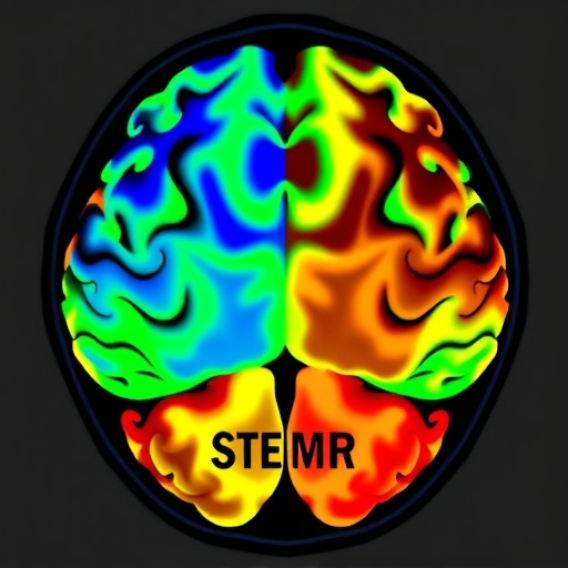
For patients with drug-resistant mesial temporal lobe epilepsy, the decision to operate hinges on a question that has long frustrated epilepsy surgeons: not just where the seizure-generating lesion sits, but what it is made of. A new study published in the European Journal of Nuclear Medicine and Molecular Imaging now shows that pairing two molecular imaging tracers in a single PET/MR session can answer both questions at once. By combining fluorine-18 fluorodeoxyglucose (18F-FDG), which maps cerebral glucose metabolism, with 18F-DPA-714, a radiotracer that binds the 18-kDa translocator protein (TSPO) expressed by activated microglia and reactive astrocytes, a team at Xuanwu Hospital of Capital Medical University in Beijing has demonstrated that the two tracers carry complementary and largely non-overlapping clinical information. TSPO imaging pinpointed the epileptogenic lesion with greater spatial precision, while FDG imaging proved superior at telling surgeons which pathological subtype they were dealing with—hippocampal sclerosis or gliosis only—a distinction with direct consequences for surgical prognosis.
The clinical stakes are considerable. Mesial temporal lobe epilepsy is the most common form of refractory focal epilepsy in adults, and surgery offers many of these patients the only realistic chance of seizure freedom. But outcomes depend heavily on the underlying pathology. Hippocampal sclerosis, characterized by progressive neuronal loss and gliosis within the hippocampal formation, is classically associated with better postoperative seizure outcomes than cases in which histopathology reveals gliosis alone, without the frank structural devastation of sclerosis. Patients whose resected tissue shows only gliosis have historically fared worse, and distinguishing between the two subtypes before surgery—when it can actually influence surgical planning—has remained difficult. Magnetic resonance imaging can be subtle or even negative, and standard visual readout of FDG-PET scans is subjective and insensitive. The Beijing team, led by corresponding author Jie Lu of the Department of Radiology and Nuclear Medicine, set out to determine whether quantitative dual-tracer imaging could fill this gap.
The study enrolled patients with unilateral refractory mesial temporal lobe epilepsy who subsequently underwent epilepsy surgery. Histopathological examination of the resected specimens allowed retrospective classification into two groups: 43 patients with hippocampal sclerosis and 41 patients with gliosis only. Crucially, each patient underwent sequential integrated PET/MR imaging with both tracers within a single week, minimizing the risk that disease evolution or metabolic fluctuation would confound comparisons between scans. The PET/MR platform matters here: by acquiring PET data simultaneously with high-resolution structural MRI, the investigators could register tracer signal precisely onto individual hippocampal and parahippocampal anatomy, enabling subregional analysis that would be difficult with standalone PET and MR. Age- and sex-matched healthy controls were recruited for comparison, and all group-level findings were subjected to false discovery rate correction for multiple comparisons.
At the whole-brain level, both patient groups showed the expected signature of mesial temporal lobe epilepsy: significant hypometabolism on FDG-PET alongside increased uptake of the TSPO tracer, with the abnormalities more pronounced in the hippocampal sclerosis group than in the gliosis-only group. But the two tracers told strikingly different stories about the spatial extent of disease. FDG hypometabolism tended to spread well beyond the temporal lobe, reflecting the fact that glucose metabolism is disturbed across widely distributed functional networks in epilepsy—changes that may represent disconnection and deafferentation of remote cortical and subcortical regions rather than the epileptogenic focus itself. In contrast, 18F-DPA-714 abnormalities remained more confined to the ipsilateral temporal lobe, providing clearer lateralization and localization of the epileptogenic lesion. The finding fits the biology: TSPO expression rises when glial cells are activated at or near the site of recurrent seizure activity, making the inflammation signal a more focal marker of the lesion than the metabolic signal.
Yet when the question shifted from where to what, the ranking reversed. In the temporal-lobe subregional analysis, FDG-PET outperformed TSPO-PET in discriminating hippocampal sclerosis from gliosis only. The best performance came from a quantitative measure: an asymmetry index computed from standardized uptake value ratios within the hippocampus. Using this hippocampal asymmetry index, the investigators correctly classified the pathological subtype in 35 of 43 patients with hippocampal sclerosis, corresponding to 81.4 percent, compared with 22 of 41 patients, or 53.7 percent, in the gliosis-only group—a statistically significant difference, with a p value of 0.007. The asymmetry index works by quantifying the left-to-right ratio of tracer uptake in homologous structures, converting what a human reader might perceive as a subtle asymmetry into an objective numeric threshold. Because hippocampal sclerosis typically produces unilateral, severe metabolic depression while gliosis-only pathology tends to be less devastating, the magnitude of metabolic asymmetry effectively encodes the underlying histology.
The study’s third major finding concerns the added value of quantification itself, independent of tracer choice. Visual inspection of PET images—the traditional mainstay of clinical presurgical evaluation—proved markedly less sensitive than quantitative analysis for both tracers. The improvement was greatest for TSPO imaging in the gliosis-only group, where localization accuracy nearly doubled, rising from 10 of 41 patients, or 24.4 percent, with visual reading to 19 of 41 patients, or 46.3 percent, with quantitative analysis. This statistically significant gain, with a p value of 0.038, is striking because gliosis-only tissue generates a weaker and more spatially circumscribed inflammatory signal than sclerotic hippocampus, precisely the situation in which a subjective eye is most likely to miss the abnormality. The asymmetry-index approach, by contrast, extracts a lateralized signal even from visually unremarkable scans.
Taken together, the results sketch a division of labor for the two tracers in the presurgical workup. TSPO-PET with 18F-DPA-714 functions as the locator: it identifies the epileptogenic zone with spatial precision grounded in the biology of neuroinflammation, and quantitative analysis substantially raises its yield in histologically mild cases. FDG-PET functions as the classifier: its metabolic fingerprints across temporal-lobe subregions, quantified through asymmetry indices, discriminate sclerotic from non-sclerotic pathology, informing expectations about surgical prognosis and potentially the extent of resection. The authors conclude that dual-tracer PET/MR combined with quantitative analysis may improve subtype-specific presurgical localization in refractory mesial temporal lobe epilepsy—an integrated assessment in which each molecular signal answers the question the other cannot.
The work builds on a rapidly evolving literature. TSPO is a mitochondrial membrane protein upregulated in activated microglia and reactive astrocytes, and radiotracers targeting it, second-generation ligands such as DPA-714 among them, have been explored for more than a decade as windows into brain inflammation. Earlier studies documented elevated TSPO binding in temporal lobe epilepsy, and subsequent work extended the approach to focal cortical dysplasia and autoimmune encephalitis, including recent demonstrations of 18F-DPA-714 PET/MRI for detecting inflammatory brain disease. Meanwhile, quantitative FDG-PET analysis has matured from simple visual asymmetry assessment into voxel-wise statistical comparisons against healthy controls, with studies showing that quantitative PET can localize lesions in MRI-negative epilepsy where conventional imaging fails. The present study is notable for combining both tracers in the same patients, in the same scanning week, on the same integrated PET/MR platform, and for validating the imaging findings against gold-standard histopathology rather than clinical outcome alone.
Several caveats temper the enthusiasm. The study was retrospective, which raises the possibility of selection bias, and the pathological classification—hippocampal sclerosis versus gliosis only—was made on resected tissue, meaning the imaging approach can only be fully validated in patients who go on to surgery. The gliosis-only category itself remains biologically heterogeneous, and recent work has highlighted “hippocampal innate inflammatory gliosis only” as a distinct entity in pharmacoresistant temporal lobe epilepsy, in which inflammatory glial changes without overt neuronal loss drive seizures. It is precisely this entity that TSPO imaging might be uniquely positioned to detect, given that the tracer’s target is inflammation itself. The investigators also note that TSPO imaging carries known limitations, including a common genetic polymorphism, the rs6971 variant, that alters tracer binding affinity and can complicate between-subject comparisons—although within-patient asymmetry measures largely sidestep this issue. Larger, prospective, multicenter studies will be needed to determine whether the reported accuracies generalize, and whether dual-tracer imaging ultimately changes surgical decision-making and seizure outcomes rather than simply refining localization statistics.
Even so, the study lands at a moment when the field is actively seeking better tools. A substantial fraction of candidates for epilepsy surgery have MRI-negative or subtly abnormal scans, and the International League Against Epilepsy has emphasized the value of multimodal neuroimaging in the presurgical workup of drug-resistant focal epilepsy. Expert consensus recommendations also urge earlier referral for surgical evaluation, heightening the need for noninvasive imaging that can characterize pathology without waiting for histology. If the Xuanwu results are confirmed, the practical pathway is straightforward: a single PET/MR session incorporating both an FDG and a TSPO tracer, analyzed with asymmetry indices derived from standardized uptake ratios, could give epilepsy surgeons a preoperative estimate of what they will find under the microscope—information that today arrives only after the operation, when it can no longer influence the plan. In a disease where the difference between two histological diagnoses can mean the difference between a seizure-free life and continued suffering, converting a post-hoc answer into a preoperative one may prove the study’s most lasting contribution.
Cite Scienmag News
Kendall Mcintyre. (September 8, 2026). Dual PET/MR tracers help classify refractory temporal lobe epilepsy subtypes. Scienmag. https://scienmag.com/dual-pet-mr-tracers-help-classify-refractory-temporal-lobe-epilepsy-subtypes/
Kendall Mcintyre. "Dual PET/MR tracers help classify refractory temporal lobe epilepsy subtypes." Scienmag, 8 September 2026, https://scienmag.com/dual-pet-mr-tracers-help-classify-refractory-temporal-lobe-epilepsy-subtypes/. Accessed 8 September 2026.
Kendall Mcintyre. "Dual PET/MR tracers help classify refractory temporal lobe epilepsy subtypes." Scienmag. September 8, 2026. https://scienmag.com/dual-pet-mr-tracers-help-classify-refractory-temporal-lobe-epilepsy-subtypes/


