


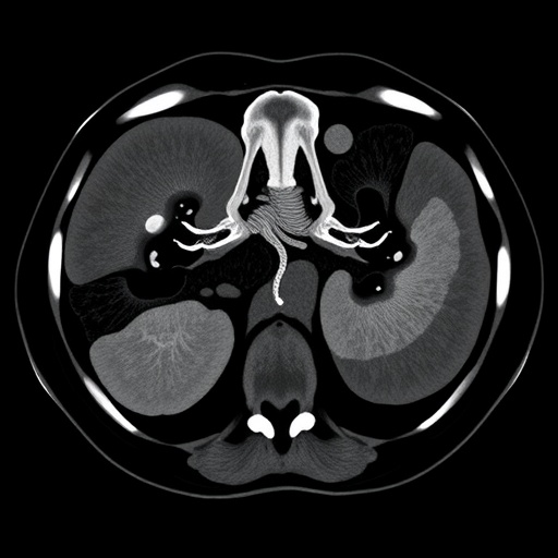
Radiologists and artificial intelligence researchers in China have unveiled a machine learning framework that can predict the aggressiveness of the most common form of kidney cancer before a patient ever sets foot in an operating room. The study, published in BMC Medical Imaging, demonstrates how a technique called radiomics—extracting vast quantities of quantitative information from ordinary medical images—can be combined with ensemble machine learning to estimate the pathological grade of clear cell renal cell carcinoma from contrast-enhanced CT scans alone.
Clear cell renal cell carcinoma, abbreviated ccRCC, accounts for the majority of kidney cancer cases worldwide, and its clinical management hinges on a pathological grading system known as WHO/ISUP grading. Under this system, pathologists examine tumor tissue under a microscope and assign a grade, typically ranging from 1 to 4, based on the prominence of nucleoli and other cellular features. The grade carries real consequences for patients: higher-grade tumors behave more aggressively, are more likely to metastasize, and influence decisions about surgery, surveillance, and adjuvant therapy. The problem is that this grading traditionally requires a biopsy or a resected specimen, meaning that oncologists and urological surgeons must often make treatment plans without knowing how aggressive the tumor actually is.
The new research, led by Dingyang Lv, Xiaomei Yao, and Jinshuai Li, with corresponding authors Ying Qiao and Weibing Shuang, set out to close that information gap. Their approach rests on a simple but powerful premise: the biological aggressiveness of a tumor leaves fingerprints in the way it enhances with contrast dye across different phases of a CT scan. Rapidly dividing, poorly differentiated tumor cells tend to have different vascular architectures, different cell densities, and different patterns of contrast wash-in and wash-out compared with indolent, well-differentiated ones. While a human radiologist can perceive some of these differences qualitatively, radiomics quantifies them at a scale and sensitivity far beyond visual assessment.
To build and test their model, the researchers assembled one of the largest cohorts yet used for this task: 884 patients with confirmed ccRCC drawn from three separate hospitals in Shanxi Province, China—the First Hospital of Shanxi Medical University, Shanxi Provincial People’s Hospital, and Shanxi Bethune Hospital. The multicenter design matters. Models trained on data from a single institution often perform impressively on that institution’s patients but collapse when confronted with scans from different CT scanners, different imaging protocols, and different patient populations. By splitting the cohort into a training set of 459 patients, an internal validation set of 198, and an external validation set of 227 drawn from other centers, the team forced their model to prove that it could generalize.
The technical pipeline began with the extraction of radiomics features from two phases of contrast-enhanced CT: the arterial phase, when contrast dye floods into the renal arteries and highlights a tumor’s blood supply, and the venous phase, when the dye has distributed more broadly through the tissue. Regions of interest were delineated around each tumor, and from these volumes the researchers computed hundreds of quantitative descriptors capturing tumor shape, first-order intensity statistics, and higher-order textures describing how pixel intensities are spatially arranged. Because raw radiomics data are notoriously noisy and redundant, the team applied feature dimensionality reduction and selection procedures, including LASSO—least absolute shrinkage and selection operator regression—to distill the raw feature set down to a stable, informative core.
What followed was an unusually systematic model-building exercise. Rather than committing to a single algorithm, the researchers fed their selected arterial-phase features, venous-phase features, and combined two-phase features into six different machine learning classifiers, generating eighteen candidate radiomics models in total. The classifier roster spanned the standard arsenal of modern machine learning: logistic regression, support vector machines, random forests, extremely randomized trees (ExtraTrees), eXtreme gradient boosting (XGBoost), and LightGBM, a gradient boosting framework known for its speed and efficiency with large feature sets. Each candidate was evaluated with receiver operating characteristic analysis, and the winner emerged clearly: an ExtraTrees model built on the combined arterial and venous phase features, denoted CTA_CTV, which achieved an area under the curve of 0.691 in the external validation set. The ExtraTrees algorithm, which injects additional randomness into how decision trees split and sample the data, has a well-earned reputation for resisting overfitting—a critical property when the number of features is large relative to the number of patients.
But the team did not stop with imaging data alone. In parallel, they used univariate and multivariate logistic regression to identify clinical and radiological characteristics—variables such as patient demographics and conventional imaging findings—that independently contributed to grading prediction, and used the ExtraTrees classifier again to construct a clinical model from these features. The final and perhaps most consequential step was fusion. Using stacking ensemble learning, a technique in which a meta-learner is trained to optimally combine the outputs of multiple base models, the researchers integrated their radiomics model and clinical model into a single decision framework, with LightGBM serving as the combiner.
The results validated the strategy. The combined model achieved an AUC of 0.703 (95% confidence interval: 0.628–0.778) in the external validation cohort, outperforming both the clinical model and the radiomics model on their own. In other words, neither the patient’s clinical picture nor the tumor’s quantitative imaging texture told the whole story alone, but stacked together they carried complementary information. Beyond raw discrimination, the team evaluated clinical usefulness using decision curve analysis, a method that quantifies the net benefit of acting on a model’s predictions across a range of decision thresholds. The combined model delivered the highest net benefit, suggesting that in practical terms it would help more patients than it would mislead.
The implications for clinical practice are substantial. Preoperative knowledge of WHO/ISUP grade could reshape how urologists counsel patients about partial versus radical nephrectomy, how they select candidates for active surveillance of small renal masses, and how they stratify risk in trials of neoadjuvant and adjuvant therapies. For patients with small kidney tumors—which are increasingly detected incidentally as abdominal imaging becomes more common—a reliable non-invasive grade estimate could mean the difference between immediate surgery and careful monitoring. The model’s modest but meaningful performance in a fully external cohort is notable in a field where reported accuracies often shrink dramatically outside the institution where the model was born.
The authors are careful to frame their findings appropriately. An AUC in the low seventies, while clinically useful, is not a substitute for pathology, and the study is retrospective in design, drawing on patients already diagnosed and treated at three centers within a single Chinese province. Broader validation across international populations, diverse scanner vendors, and prospectively collected cohorts will be needed before such models can be deployed at the bedside. The written informed consent of patients was waived owing to the retrospective nature of the work, but the study complied with the Declaration of Helsinki and received approval from the ethics boards of all three participating hospitals.
Nevertheless, the study adds to a rapidly growing body of evidence that the information needed to characterize a tumor’s biology is already embedded in images clinicians acquire every day. By demonstrating that multi-phase CT radiomics, carefully selected and intelligently fused with clinical variables, can approximate pathological grading non-invasively, the researchers have taken a concrete step toward a future in which risk stratification for kidney cancer begins the moment a scan is performed—before a single cell has been examined under a microscope. The work was published open access, and the authors declare no competing interests and received no external funding for the study.
Cite Scienmag News
Nathaniel Bowman. (September 7, 2026). Multicenter study predicts kidney cancer grade using multi-phase CT radiomics. Scienmag. https://scienmag.com/multicenter-study-predicts-kidney-cancer-grade-using-multi-phase-ct-radiomics/
Nathaniel Bowman. "Multicenter study predicts kidney cancer grade using multi-phase CT radiomics." Scienmag, 7 September 2026, https://scienmag.com/multicenter-study-predicts-kidney-cancer-grade-using-multi-phase-ct-radiomics/. Accessed 7 September 2026.
Nathaniel Bowman. "Multicenter study predicts kidney cancer grade using multi-phase CT radiomics." Scienmag. September 7, 2026. https://scienmag.com/multicenter-study-predicts-kidney-cancer-grade-using-multi-phase-ct-radiomics/


