


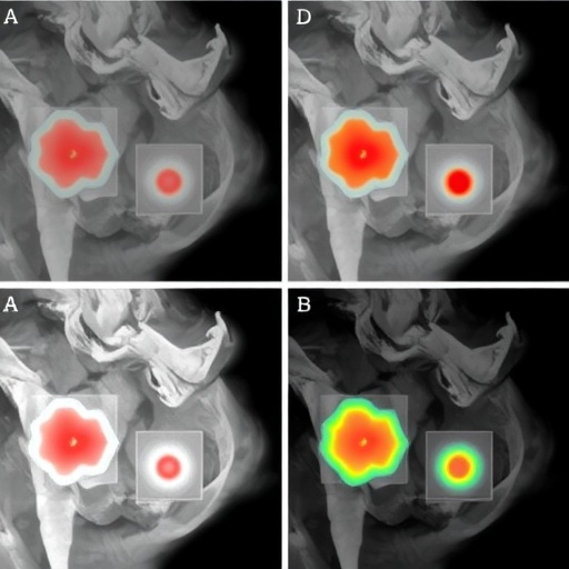
Melanoma, the deadliest form of skin cancer, often presents as a subtle dark patch on the skin whose edges blur almost imperceptibly into healthy tissue. Detecting those edges automatically is one of the deceptively hard problems in medical image analysis, and a new open-access study now offers an unusually elegant answer: instead of forcing a neural network to decide pixel by pixel whether something is “lesion” or “not lesion,” the researchers behind a new architecture called FAED let the network think in shades of uncertainty — the way a dermatologist actually does.
The work, published in the journal Complex & Intelligent Systems, comes from an international team spanning SRM Institute of Science and Technology in India, the National Institute of Technology Rourkela, China University of Mining and Technology, Innopolis University in Russia, and St. Petersburg Electrotechnical University “LETI.” The team — M. R. Indresh, Soumyajit Gayen, Dmitrii Minenkov, Dmitrii Kaplun and Ram Sarkar — describes FAED, a Fuzzy Attention-aided Encoder-Decoder architecture, which swaps out the rigid binary logic of standard attention mechanisms for a soft, probabilistic notion of relevance inspired by fuzzy set theory. The results are striking not only for their accuracy but for the architecture’s remarkable frugality: with just 2.4 million parameters and roughly 4 GFLOPs of computation, FAED posts Dice scores that put it at the top tier of contemporary segmentation models while running at inference speeds measured in milliseconds.
The clinical stakes of this problem are easy to underestimate. Early-stage melanoma is highly curable, but the first line of defense is visual inspection of pigmented lesions, typically through dermoscopy — the imaging of skin through a magnifying device that reveals subsurface structures. Automated segmentation of dermoscopy images, the task of drawing an accurate boundary around a lesion, underpins every downstream measurement clinicians and computer-aided diagnosis systems rely on, including the asymmetry, border irregularity and color variation criteria used in melanoma risk scoring. Yet the task is plagued by low contrast between lesion and healthy skin, hair occlusions, specular reflections, and most fundamentally, ambiguous boundaries where the lesion fades gradually into its surroundings.
For years, the dominant tool for this job has been U-Net, a convolutional encoder-decoder architecture in which a contracting path extracts increasingly abstract features and an expanding path reconstructs a pixel-level prediction. The critical link between the two halves is a set of skip connections that pass fine-grained spatial detail from early encoder layers directly to the decoder. Most modern variants bolt attention modules onto these skip connections: the network learns to “gate” which features to pass through. But those gates are typically binary — a feature channel or spatial position is either kept or discarded. The FAED authors argue that this all-or-nothing logic is fundamentally mismatched to the nature of skin lesions, where the transition between sick and healthy tissue is gradual, not sharp. A binary gate discards exactly the soft, intermediate evidence that defines an ambiguous boundary.
FAED’s central innovation is its Boundary-conditioned Soft Fuzzy Attention (BSFA) module, which replaces standard skip connections altogether. Rather than multiplying features by a learned 0-or-1 mask, BSFA evaluates feature relevance using learnable Gaussian membership functions — mathematical constructs from fuzzy logic that assign each feature a continuous degree of membership, modeled as a probability-like value between zero and one. In practice, this means the network can express that a feature is “somewhat relevant” or “mostly relevant,” preserving graded boundary information that binary attention would crush. The Gaussian membership functions are themselves learnable parameters, so the network discovers its own notions of partial relevance during training rather than having them imposed by a fixed rule.
The architecture adds two further refinements that the authors show are individually and jointly important. The first is an Adaptive Fuzzy Mixture-based aggregation scheme. Features extracted at different depths of the network vary enormously in scale and semantics — shallow layers carry edge textures, deep layers carry abstract shape information — and fusing them well is a persistent headache in segmentation design. The fuzzy mixture approach treats each feature source as contributing to a soft ensemble, weighting its contribution according to a learned similarity-based membership rather than simple concatenation. The second refinement is an explicit Boundary Cue, a signal fed into the attention mechanism that modulates its focus along lesion perimeters. Where the fuzzy membership decides “how relevant” a feature is, the boundary cue tells the attention “where to look,” sharpening the model’s sensitivity precisely at the lesion border where errors are most costly.
The authors validated FAED on the four most widely used benchmarks in the field: the ISIC2016, ISIC2017 and ISIC2018 dermoscopy datasets from the International Skin Imaging Collaboration, and the smaller PH² dataset of melanocytic lesion images. The segmentation quality was measured with the Dice score, a standard metric that quantifies the overlap between the predicted lesion mask and the ground truth, where a score of 1.0 means perfect agreement. FAED achieved a Dice score of 0.9140 on ISIC2016, 0.9135 on PH², 0.8781 on ISIC2018, and 0.8615 on ISIC2017 — competitive-to-leading figures given the architecture’s size. Notably, the ISIC2016 and PH² results hover around the 0.91 mark, a level of overlap that corresponds to clinically meaningful boundary fidelity.
Just as important as the headline numbers is the efficiency profile, which the team documented with careful empirical measurements on an NVIDIA Tesla T4 GPU. FAED performs inference in 10.05 milliseconds per image at batch size 1, and 6.76 milliseconds per image when batched at 8 — throughput fast enough for real-time clinical workflows. Peak GPU memory during inference is similarly modest: 505 MB at batch size 1 and 948 MB at batch size 8. For context, many state-of-the-art segmentation models rely on heavyweight transformer backbones or large convolutional stacks with parameter counts in the tens of millions, demanding memory and compute budgets that make deployment on hospital hardware, edge devices or low-resource settings difficult. FAED’s 2.4 million parameters and roughly 4 GFLOPs place it in a different class entirely, suggesting that careful architectural design — rather than brute-force scale — can carry segmentation performance a long way.
To verify that each component of the design actually earns its place, the researchers conducted ablation studies, the standard experimental practice of removing parts of a system one at a time and measuring the drop in performance. These studies confirmed that both the prototype-based fuzzy aggregation and the boundary-conditioned modulation of attention contribute measurably to the observed improvements. In other words, the gains are not an artifact of added capacity or incidental tuning: the soft membership modeling and the explicit boundary guidance are doing real, distinguishable work. That finding matters for the broader field, because it offers evidence that how features are fused — treating fusion as a soft similarity-based membership problem — can be as consequential as how features are extracted.
The philosophical shift at the heart of FAED is worth dwelling on. Classical computer vision and early deep learning systems were built on crisp logic: a pixel belongs to a class, a feature passes a gate, a decision is yes or no. Fuzzy logic, introduced decades ago as a formal way of reasoning with degrees of truth, has long been touted as a natural fit for medical imaging, where human experts themselves reason in gradients — “this border looks slightly irregular,” “this region is probably part of the lesion.” What has changed recently is that learnable fuzzy components, such as Gaussian membership functions optimized end-to-end by gradient descent, can now be embedded inside deep networks so that the fuzzy rules themselves are discovered from data. FAED is a concrete demonstration that this marriage of classical soft-computing theory and modern deep learning can outperform hard-gated alternatives on a real clinical task, without any increase in architectural complexity.
The implications for melanoma screening are potentially significant, particularly for parts of the world where dermatologists are scarce and mobile screening programs depend on lightweight, fast and reliable algorithms. A model that runs in a few milliseconds on an entry-level GPU, fits comfortably in under a gigabyte of memory, and still achieves over 91 percent overlap with expert-annotated boundaries on benchmark datasets is precisely the kind of tool that can be embedded into telemedicine pipelines or portable dermoscope accessories. The authors caution, as all careful researchers do, that benchmark performance is a step toward clinical deployment, not the deployment itself — prospective validation on diverse skin tones, imaging devices and real-world lesion appearances remains an essential next stage for any segmentation technology destined for the clinic.
The article was published open access under a Creative Commons license, making the full technical description freely available to researchers and clinicians worldwide. The study was supported by the Ministry of Economic Development of the Russian Federation. As peer-reviewed, citable research made available early for faster dissemination, it joins a growing body of work arguing that the future of medical AI lies not only in ever-larger models, but in smarter ones — systems that, like the physicians they assist, know how to say “maybe.” FAED’s fuzzy attention may be an early but compelling example of that principle turned into working code.
Cite Scienmag News
Nathaniel Bowman. (September 3, 2026). Fuzzy attention-based encoder-decoder improves skin lesion segmentation accuracy. Scienmag. https://scienmag.com/fuzzy-attention-based-encoder-decoder-improves-skin-lesion-segmentation-accuracy/
Nathaniel Bowman. "Fuzzy attention-based encoder-decoder improves skin lesion segmentation accuracy." Scienmag, 3 September 2026, https://scienmag.com/fuzzy-attention-based-encoder-decoder-improves-skin-lesion-segmentation-accuracy/. Accessed 11 September 2026.
Nathaniel Bowman. "Fuzzy attention-based encoder-decoder improves skin lesion segmentation accuracy." Scienmag. September 3, 2026. https://scienmag.com/fuzzy-attention-based-encoder-decoder-improves-skin-lesion-segmentation-accuracy/


