


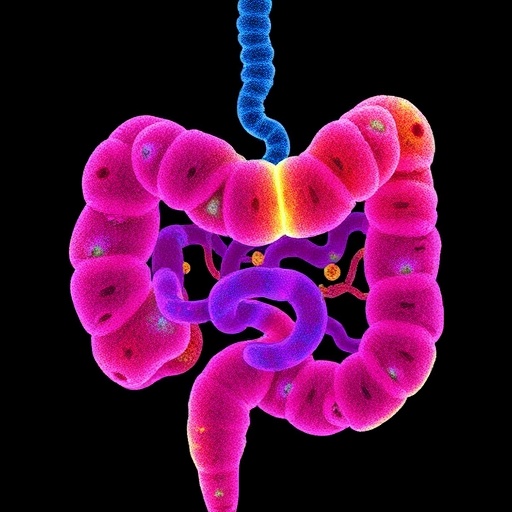
Beneath every square centimeter of the human small intestine lies a hidden scaffold of cells that has quietly governed one of the most dynamic tissues in the body. While the gut’s epithelial lining — with its rapidly cycling stem cells, mucus-secreting goblet cells, hormone-producing enteroendocrine cells and nutrient-absorbing enterocytes — has long dominated the scientific spotlight, the connective tissue immediately beneath it, known as the mesenchyme, has remained poorly charted. Now, in a study published in August 2026 in Nature Cell Biology, researchers led by K. F. Johnson, X. Dong and Y.-H. Tsai, together with their colleagues, have produced one of the most detailed maps to date of this overlooked layer, cataloguing the diversity of mesenchymal cells in the native human small intestine and comparing that landscape with the mesenchyme found in laboratory-grown intestinal organoids. Their results reveal that the gut’s supporting cells are far from an interchangeable backdrop: they form a finely patterned mosaic in which specialized populations occupy precise locations and send distinct instructions to the stem cells above them.
To appreciate why this map matters, it helps to consider the extraordinary biology of the small intestine. Its inner surface is arranged into millions of finger-like villi, each ringed at its base by pocket-like crypts that harbor stem cells marked by the receptor LGR5. These stem cells divide roughly every day, and their daughters stream upward along the villus, maturing into the specialized cells that digest food and absorb nutrients before being shed into the lumen within just a few days. Coordinating this perpetual renovation are gradients of signaling molecules. Wnt and R-spondin signals, acting through LGR5 and its partners RNF43 and ZNRF3, keep stem cells proliferating at the crypt base, while bone morphogenetic protein, or BMP, signaling near the villus tip nudges their descendants toward mature, specialized fates. Crucially, many of these instructive signals are not produced by the epithelium itself but by the mesenchyme beneath it, which secretes Wnt ligands, R-spondins, BMP antagonists such as gremlin 1, and growth factors such as HGF that together sustain the stem cell niche.
Despite this central role, the human intestinal mesenchyme has been notoriously difficult to study. Its cells look structurally similar under the microscope, lack easily readable surface markers, and are embedded in a dense meshwork of muscle, nerve and blood vessels that complicates their isolation. Johnson, Dong, Tsai and their colleagues tackled the problem by combining single-cell RNA sequencing with spatial mapping. In droplet-based single-cell transcriptomics, thousands of individual cells are captured in tiny water-in-oil droplets, and the messenger RNA within each cell is barcoded and sequenced, allowing every cell’s gene-expression profile to be read out and then grouped by computational clustering into distinct types and states. The team profiled mesenchymal cells isolated from human small intestinal tissue, enriching for stromal populations and setting aside immune and epithelial cells, and then used spatial techniques, which preserve the physical position of each cell, to anchor those expression profiles back to their anatomical homes along the crypt-villus axis and across the gut wall. The same strategy was applied to intestinal organoids, the self-organizing mini-guts grown in three-dimensional matrices that have become a mainstay of intestinal research.
The resulting atlas paints a picture of unexpected richness. Rather than a uniform population of fibroblasts, the human small intestinal mesenchyme resolved into a broad collection of transcriptionally distinct populations, each defined by its own combination of marker genes and secreted signals. Among them were subepithelial myofibroblasts — contractile cells marked by the gene ACTA2 that form a sheath immediately beneath the epithelium; fibroblast populations enriched for the Wnt-potentiating molecule RSPO3 and for the BMP antagonist GREM1; long, slender telocyte-like cells marked by FOXL1 that extend thin processes around crypt structures; pericytes marked by PDGFRB and RGS5 that embrace blood vessels and help regulate local blood flow; and fibroblast populations associated with the smooth muscle layers deeper in the gut wall. Strikingly, each population occupied a stereotyped position: some sheathed the crypt base, others lined the villus core, and others tracked the vasculature or muscle, giving the mesenchyme a reproducible anatomical architecture.
The spatial arrangement of these cells is anything but random, and it maps cleanly onto the signaling logic of the stem cell niche. Mesenchymal cells lying close to the crypt base carried the ligands that activate Wnt signaling and the antagonists that suppress BMP activity, creating a permissive zone in which stem cells can maintain themselves indefinitely. Farther up the villus the balance flips: mesenchymal cells in the villus core express BMP ligands, reinforcing the differentiation program of epithelial cells on their way to the tip. The epithelium, in turn, talks back. Differentiating epithelial cells emit Indian hedgehog, which is received by the underlying mesenchyme and helps pattern fibroblast identity and positioning, closing a reciprocal loop between the two tissue layers. Taken together, the findings suggest that the niche is best understood not as a single cell type or a single molecule but as a geometric arrangement of specialized supporting cells whose locations, and the signals concentrated at those locations, encode the intestinal blueprint.
The comparison with organoids is where the study delivers its most sobering message. Standard human intestinal organoids, grown in three-dimensional matrices and bathed in culture medium containing exogenous Wnt, R-spondin, Noggin and EGF, are essentially pure epithelium: they recapitulate crypt-villus architecture in miniature but contain no mesenchymal cells at all, which is precisely why their culture medium must supply the niche signals that the mesenchyme normally provides. Organoids that do include mesenchyme, such as human intestinal organoids derived from pluripotent stem cells that spontaneously develop both epithelial and mesenchymal compartments, come closer to the real thing. Yet the new atlas shows that even these models fail to capture the full spectrum of mesenchymal diversity seen in living tissue. Certain subtypes are under-represented or absent altogether, and the proportions of others are skewed, meaning that organoid-based experiments may systematically miss mesenchyme-dependent biology, from niche signaling to interactions with immune and vascular cells.
These gaps have practical consequences. Because organoids are increasingly used to test drugs, model disease and probe gene function, a model lacking key supporting cells may respond differently from the tissue it is meant to represent. The atlas therefore provides both a reference against which organoid fidelity can be measured and a parts list for rebuilding it. Knowing which mesenchymal subtypes supply which signals gives researchers a rational basis for engineering improved organoids, for example by co-culturing defined fibroblast populations with epithelium, by tuning culture conditions to preserve fragile subtypes, or by adding recombinant versions of the ligands that organoid mesenchyme fails to produce. The map also offers clues about injury and repair: studies across the field have shown that when the intestinal epithelium is wounded, nearby mesenchymal cells re-enter developmental-like states and ramp up niche signals to drive regeneration, and a complete inventory of resting states makes it easier to identify and study these activated ones.
The clinical implications extend well beyond the culture dish. The small intestine is the primary battleground of Crohn’s disease, and mesenchymal cells are central both to its repair responses and to its most feared complication, fibrosis, in which activated myofibroblasts lay down scar tissue that narrows the bowel and often forces patients into repeated surgery. The new map identifies the normal states against which disease-activated fibroblast populations can be compared, and it highlights the communication channels that mesenchymal cells use to talk to epithelium, immune cells and blood vessels — channels that could, in principle, be targeted to dampen inflammation or limit scarring. Mesenchymal biology is also implicated in radiation injury to the gut, in the intestine’s failure to adapt after massive surgical resection, and in cancers of the gastrointestinal tract, where tumor-associated fibroblasts are known to hijack and corrupt normal niche programs to support malignant growth.
The study also underscores how much can be lost when findings in mice are extrapolated to humans. Much of what is known about the intestinal niche was worked out in mouse models, and while many mesenchymal programs are conserved across species, the human map brought the corresponding human cell types and their markers into sharp focus, reinforcing the value of building comprehensive atlases directly in human tissue. It also strengthens the case for organoids as a human-relevant experimental platform: if researchers know exactly which mesenchymal cells are missing, they can add them back, turning a simplified model into one that reflects human anatomy and physiology more faithfully. In this sense the work joins a broader scientific effort, exemplified by initiatives such as the Human Cell Atlas, to replace simplified diagrams of organ biology with dense, multidimensional maps of the actual cells involved, their positions and their conversations.
Looking forward, the atlas is likely to become a community resource for anyone attempting to grow gut tissue that behaves like the real thing. Tissue-engineered intestine, built from stem cells and biomaterial scaffolds in the laboratory and ultimately intended for patients with intestinal failure, will need a correctly patterned mesenchyme if it is to develop functional crypts and villi, and the new map supplies something close to a specification sheet. The same reference should accelerate organoid platforms for personalized medicine, in which a patient’s own cells are used to model their disease and predict their response to therapy with the niche fully represented. For a layer of the gut long treated as background scenery, the mesenchyme has now been given its own detailed portrait, and the portrait makes one thing clear: the small intestine’s celebrated stem cells are only half the story. The other half lives just beneath them — patterned, specialized and indispensable.
Cite Scienmag News
Drew Townsend. (August 30, 2026). New atlas reveals mesenchymal diversity in human gut and organoids. Scienmag. https://scienmag.com/new-atlas-reveals-mesenchymal-diversity-in-human-gut-and-organoids/
Drew Townsend. "New atlas reveals mesenchymal diversity in human gut and organoids." Scienmag, 30 August 2026, https://scienmag.com/new-atlas-reveals-mesenchymal-diversity-in-human-gut-and-organoids/. Accessed 30 August 2026.
Drew Townsend. "New atlas reveals mesenchymal diversity in human gut and organoids." Scienmag. August 30, 2026. https://scienmag.com/new-atlas-reveals-mesenchymal-diversity-in-human-gut-and-organoids/


