

An AI Network Maps the Eye’s Tiny Blood Vessels to Reveal Hidden Signs of Disease
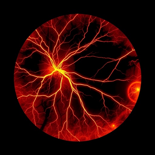
A new artificial-intelligence system could turn the branching blood vessels visible at the back of the eye into a more precise source of medical information. Called MATHFI, the system analyzes retinal photographs and separates blood vessels from surrounding tissue, including the finest branches that are often difficult for automated methods to detect. In a study published in the Journal of Medical and Biological Engineering, researchers report that the network preserved the structure and continuity of retinal vessels while reducing false signals caused by the optic disc, bright lesions, uneven illumination, and other anatomical features. The goal is not simply to create a cleaner image. By producing a more faithful map of the retinal circulation, MATHFI is designed to support quantitative measurements that may help characterize diabetic retinopathy and other disorders linked to vascular damage.
The retina offers a rare view of the body’s microcirculation without the need for surgery. Light entering the eye is focused onto a layer of neural tissue supplied by a dense network of arteries, veins, and capillaries. In a standard fundus photograph, these vessels appear as dark, branching structures against a brighter background. Their arrangement, width, curvature, density, and branching complexity can change with disease. Yet extracting those features computationally is challenging. Thin vessels may occupy only a few pixels, contrast can vary across an image, and lesions or anatomical boundaries may resemble vessel segments. A model that incorrectly fills in background regions or breaks a vessel into disconnected pieces can distort downstream measurements, potentially making a healthy vascular network appear abnormal or masking a real change.
MATHFI addresses the problem by combining several image-processing and deep-learning strategies in one pipeline. Its name refers to Multi-scale Adaptive Thresholding with Hierarchical Feature Integration. Adaptive thresholding estimates whether a pixel belongs to a vessel relative to its local surroundings rather than applying one fixed brightness cutoff to the entire photograph. This is important because illumination is rarely uniform across a fundus image. The network also examines information at multiple spatial scales, allowing it to recognize both broad vessels and narrow peripheral branches. In effect, the model must solve two related problems at once: identifying the local visual signature of a vessel and understanding whether that candidate segment fits into the larger vascular architecture.
The system’s neural architecture uses attention-guided feature modulation, hierarchical skip fusion, multi-scale subtraction, adaptive selective fusion, and a final refinement stage. These terms describe ways of preserving useful information as an image passes through the network. In an encoder-decoder model, the encoder progressively compresses an image to learn increasingly abstract features, while the decoder reconstructs a pixel-level segmentation map. Skip connections transfer high-resolution details from early layers to later reconstruction stages, helping recover vessel boundaries that might otherwise be lost. MATHFI’s hierarchical fusion combines features from different depths rather than treating them as interchangeable. Attention mechanisms further assign greater weight to image regions and features that appear relevant to vessels, while suppressing patterns more likely to represent background anatomy or imaging artifacts.
Topology is central to the researchers’ approach. A vessel map is not merely a collection of correctly labeled pixels; it is also a network of connected paths. If a narrow vessel is interrupted by even a small gap, measurements of branching, tortuosity, or network density can change substantially. To assess this structural property, the study used clDice, a topology-aware metric that evaluates the overlap between the predicted and reference vessel skeletons as well as their regions. Conventional measures such as sensitivity and specificity count correctly classified pixels, but they may not adequately penalize a model that produces a visually plausible yet disconnected network. By emphasizing centerline continuity, clDice is better suited to the biological geometry of vessels, where connectivity matters for both interpretation and quantitative analysis.
The researchers trained and evaluated MATHFI on three publicly available retinal image datasets: DRIVE, CHASE-DB1, and STARE. These collections differ in image characteristics and acquisition conditions, making them useful benchmarks for testing whether a model can cope with variation beyond a single source. Using Monte Carlo cross-validation, the team repeatedly divided the data for training and evaluation rather than relying on one fixed split. On DRIVE, MATHFI achieved a sensitivity of 83.06 ± 0.92, a specificity of 98.09 ± 0.13, and a clDice score of 81.81 ± 0.36. On CHASE-DB1, the reported values were 85.00 ± 1.89 for sensitivity, 98.59 ± 0.24 for specificity, and 84.60 ± 0.66 for clDice. On STARE, the system reached 80.68 ± 8.49 sensitivity, 98.32 ± 0.61 specificity, and 82.65 ± 3.69 clDice. The variation in the results is itself informative: performance was less consistent on STARE, suggesting that image diversity remains a meaningful challenge.
The study then used MATHFI-generated vessel masks for a separate analysis of diabetic retinopathy. The researchers examined images from the APTOS 2019 Blindness Detection dataset, which includes five severity groups: no diabetic retinopathy, mild, moderate, severe, and proliferative disease. Rather than limiting the analysis to whether a vessel was present, they calculated four vascular biomarkers: fractal dimension, vascular density, tortuosity, and caliber. Fractal dimension is a mathematical description of how completely a branching pattern occupies space; in retinal imaging, it can capture aspects of the complexity of the vascular tree. Vascular density estimates the proportion or amount of vessel structure in a defined region. Tortuosity measures how much vessels deviate from a straighter course, while caliber describes their apparent width. Each measure provides a different view of retinal architecture, and each can be evaluated globally, within rings around the optic disc, or across the superior, inferior, nasal, and temporal regions represented by ISNT quadrants.
Across the diabetic retinopathy groups, the researchers found significant differences in all four biomarkers using one-way analysis of variance, with p values below 0.001. The reported global pattern included declining fractal dimension and vascular density as disease severity increased. Such a trend is biologically plausible because progressive retinal damage can involve capillary loss, nonperfusion, and remodeling of the vascular network, although the exact appearance of disease may differ between patients and anatomical regions. Changes in tortuosity and caliber can reflect vessel stress, abnormal remodeling, or altered blood flow, but these measurements are sensitive to image quality and segmentation errors. The researchers therefore present the biomarker results as evidence that the segmentation framework can enable quantitative analysis, not as proof that MATHFI alone can diagnose or stage an individual patient.
That distinction is important as artificial intelligence moves from experimental image analysis toward clinical decision support. A high-performing segmentation model does not automatically become a validated screening tool. It must be tested on diverse populations, cameras, image resolutions, ethnic groups, disease profiles, and real-world clinical workflows. It also needs to be compared with expert graders and evaluated for failure modes, including poor focus, media opacity, unusual anatomy, hemorrhages, exudates, and images captured outside the training distribution. The MATHFI study used established public datasets and reported an ablation analysis to examine the contributions of its major components, but the abstract does not establish prospective clinical performance or demonstrate that the system improves patient outcomes. Its immediate contribution is more foundational: a method for generating vessel maps that retain fine structures and support reproducible measurements.
If the approach withstands broader validation, its potential reach could extend beyond diabetic retinopathy. Retinal vascular patterns have been investigated in connection with hypertension, glaucoma, choroidal disease, and systemic vascular conditions. A reliable segmentation layer could allow researchers to measure subtle changes over time, compare vascular regions within the same eye, and combine vessel-derived features with lesion detection or other clinical signals. It could also make automated analysis more useful in settings where specialist graders are scarce, provided that appropriate safeguards and human oversight are maintained. For now, MATHFI is best understood as an enabling technology rather than a replacement for ophthalmic expertise. By treating the retina’s vessels as a connected biological network instead of a set of isolated dark pixels, the system points toward a future in which a routine eye photograph yields a richer, more quantitative portrait of disease.
Cite this page
SCIENMAG. (August 27, 2026). AI Network Sharpens Retinal Vessel Mapping and Biomarker Analysis Across Scales. https://scienmag.com/ai-network-sharpens-retinal-vessel-mapping-and-biomarker-analysis-across-scales/
SCIENMAG. "AI Network Sharpens Retinal Vessel Mapping and Biomarker Analysis Across Scales." Scienmag, 27 August 2026, https://scienmag.com/ai-network-sharpens-retinal-vessel-mapping-and-biomarker-analysis-across-scales/. Accessed 27 August 2026.
SCIENMAG. "AI Network Sharpens Retinal Vessel Mapping and Biomarker Analysis Across Scales." Scienmag. August 27, 2026. https://scienmag.com/ai-network-sharpens-retinal-vessel-mapping-and-biomarker-analysis-across-scales/


