


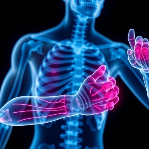
Muscle and bone are often discussed as separate systems: one powers movement, while the other forms the body’s structural framework. Yet they develop, adapt and deteriorate in close relationship. A large analysis of UK Biobank data now suggests that sarcopenia—the loss of skeletal-muscle mass, strength and function—and osteoporosis may be reciprocal risk factors, with each condition associated with a higher likelihood of the other. Their shared biology appears to involve genetic variation, immune signalling, inflammation and metabolism. Together, the findings support the idea of osteosarcopenia, a combined decline in muscle and bone health, as more than the simple coexistence of two age-related disorders. The researchers analysed data from more than 500,000 adults aged 40 to 69 at UK Biobank’s baseline assessment. They examined appendicular lean mass adjusted for height squared, hand-grip strength and usual walking pace as measures related to sarcopenia. Osteoporosis was identified through linked health and primary-care records using ICD-10 codes M80–M81, while bone mineral density (BMD) was assessed mainly at the heel using quantitative ultrasound. The team combined epidemiological analysis with genome-wide association studies, Mendelian randomisation, plasma proteomics, metabolomics, single-cell RNA sequencing and pathway analysis.
The clearest longitudinal signal ran from muscle traits to later osteoporosis. Participants with higher appendicular lean mass, stronger hand grips or faster usual walking speeds had lower observed risks of developing osteoporosis. For each standardised increase in the three measures, the reported hazard ratios were 0.830 for adjusted lean mass, 0.544 for grip strength and 0.735 for walking pace. A hazard ratio below one indicates a lower event rate in the model, although it does not by itself prove that improving the trait would prevent disease. When the three muscle measures were adjusted for one another, grip strength and walking pace remained independently associated with lower osteoporosis risk, with hazard ratios of 0.593 and 0.788. The exposure–response patterns were broadly monotonic for these functional measures: risk fell as strength and walking speed increased. Analyses also detected interactions with age, sex and body-mass index. The associations were generally stronger in younger adults; lean mass and walking pace had larger effects in men, while lean mass showed a stronger relationship among people with lower BMI. Even within these subgroups, however, favourable muscle traits were consistently associated with lower risk.
Muscle mass itself produced a less straightforward result. Adjusted appendicular lean mass showed a U-shaped association with osteoporosis, meaning that neither the lowest nor the highest levels appeared optimal in the statistical model. This finding is exploratory and does not demonstrate that unusually high muscle mass causes bone loss. The authors propose several possible explanations that require testing, including overtraining, inadequate energy availability and exercise-related hormonal disruption. Muscles normally load the skeleton through contraction, encouraging bone adaptation, but that response may reach a ceiling. Prolonged fatigue, irregular forces or abnormal stress distribution could alter the benefit of loading and contribute to skeletal microdamage. Intense training combined with low energy availability has also been linked to impaired bone health, while hormonal disruption may add to the risk. These possibilities should not be read as evidence that ordinary physical activity is harmful. Rather, the unusual curve highlights the limitations of interpreting a single measure of lean mass without detailed information about training intensity, nutrition, hormones, body composition and previous injuries. Replication in other populations will be needed before the pattern can influence advice or clinical practice.
The association also ran in the opposite direction. In longitudinal analyses, people with higher left- or right-heel BMD, or higher heel BMD T-scores, were less likely to develop probable sarcopenia. The relationship was broadly monotonic: as heel density rose, sarcopenia risk fell. Complementary analyses using BMD measured at the femoral neck and lumbar spine by dual-energy X-ray absorptiometry supported inverse associations with both probable sarcopenia and confirmed sarcopenia. The primary sarcopenia definition was “probable sarcopenia”, based on low grip strength alone—below 27 kilograms in men or 16 kilograms in women—while confirmed sarcopenia additionally required low adjusted appendicular lean mass. Genetic analyses added evidence of two-way effects. Mendelian randomisation indicated that most sarcopenia-related traits, but not usual walking pace, had effects on lumbar-spine BMD. In the reverse analysis, lumbar-spine BMD showed effects on most sarcopenia traits, again excluding walking pace. Mendelian randomisation uses genetic variants as instruments to reduce some confounding, but its validity depends on assumptions about those variants. It is not a substitute for a randomised intervention trial.
The investigators next searched for circulating molecules that might help explain how muscle status relates to bone. Mediation analysis identified 465 proteins associated with the lean-mass measure, 706 associated with grip strength and 697 associated with walking pace in pathways leading statistically towards osteoporosis risk. Of these, 352 were shared across all three muscle traits. The metabolite analysis identified 21 potential mediators for lean mass, 140 for grip strength and 127 for walking pace, including 17 shared across the three measures. Because exposure, mediator and outcome were not measured repeatedly in a way that established their temporal order, these results indicate statistical mediation consistent with a biological pathway, not proof that a particular protein or metabolite causes muscle loss to produce osteoporosis. To investigate where the proteins might arise, the researchers compared their signals with single-cell RNA-sequencing data from 90,902 cells taken from intercostal muscles of 17 donors in a human skeletal-muscle ageing atlas. Mediator-related expression was especially enriched in myeloid, endothelial and stromal cells. These cell groups participate in immune regulation, blood-vessel biology and connective-tissue maintenance, suggesting that muscle-to-bone communication may involve secreted signals and local cellular networks as well as mechanical loading.
Genetic and biomarker analyses pointed to a common background for the two disorders. Across the genome, the estimated genetic correlation between sarcopenia and osteoporosis risk was 0.25. Local analysis identified 102 genomic regions with significant heritability for both traits; 12 also showed significant local genetic correlation, and 10 of those correlations were positive. Polygenic risk scores provided a similar pattern: a higher genetic risk score for sarcopenia was associated with osteoporosis prevalence and incidence, while an osteoporosis score was associated with both the prevalence and incidence of sarcopenia. Circulating biomarkers overlapped extensively as well. Of 226 metabolites associated with sarcopenia, 164—71 per cent—were also associated with osteoporosis, all in the same direction. Among 1,576 sarcopenia-associated proteins, 502—31.3 per cent—were also linked to osteoporosis; 500 had concordant directions of association. Most of the shared proteins were secreted molecules whose gene activity was enriched in immune and lymphoid tissues. A protein–protein interaction network was far more connected than expected by chance, with immune-regulatory nodes including IL6, CXCL8 and TNF. These are associations, not diagnostic markers or validated treatment targets.
The biological pathways highlighted by the analysis offer a plausible bridge between muscle and bone. Enriched pathways included T-helper-17-cell differentiation, interleukin-17 signalling and NF-κB signalling. Chronic inflammatory activity can promote muscle protein breakdown and atrophy, while inflammatory signalling in bone can stimulate osteoclasts, the cells responsible for bone resorption. Several regulatory factors identified in the analysis have established roles across these processes, including NFKB1, RELA, FOXO1, SIRT1 and the vitamin D receptor. Other candidates included TFAM, which is involved in mitochondrial DNA maintenance; MGP, a vitamin K-dependent protein involved in mineral regulation and also linked to muscle development; and COMMD7, which has been connected with NF-κB activity. However, the gene-expression results demand caution. Transcriptome-wide association and summary-data Mendelian-randomisation analyses found dozens of genes nominally associated with both conditions in several tissues, and about 70 per cent showed consistent effect directions. After correction for multiple testing, no single gene remained significantly associated with both diseases simultaneously. The authors therefore describe these findings as exploratory clues rather than confirmed mechanisms. Similar caution applies to the large lists of proteins and metabolites: shared statistical signals need experimental validation.
The study also found common patterns for several potentially modifiable factors. Greater adult smoking exposure, measured in pack-years, was associated with higher prevalence of both sarcopenia and osteoporosis. Sleep duration showed a U-shaped relationship: sleeping fewer than six hours or more than eight hours was associated with greater risk than sleeping six to eight hours. More physical activity was associated with lower prevalence of both disorders up to a point, after which the association was no longer statistically significant. These observations do not establish that changing sleep duration or activity to a particular level will prevent either condition, and the study cannot distinguish all the reasons why people sleep or exercise more or less. Still, the molecular analyses found substantial overlap in the proteins and metabolites associated with these exposures and both diseases—roughly 30.5 to 73 per cent depending on the factor and analysis. Smoking and short sleep shared mediators linked to inflammation, oxidative stress, lipid metabolism and gastrointestinal function. Physical activity shared metabolic mediators, particularly involving lipid metabolism. The patterns are consistent with a muscle–bone unit influenced by systemic inflammation and metabolic health, while supporting established preventive priorities such as avoiding tobacco and maintaining appropriate physical activity and sleep.
Several limitations define what can be concluded. The primary sarcopenia outcome relied on grip strength alone, which may misclassify people whose muscle mass or mobility differs from their hand strength. The timing of sarcopenia onset was also uncertain; people who developed it between the baseline and imaging visits may have been excluded from some analyses because of progression or death. Heel BMD, the main osteoporosis trait, was estimated with quantitative ultrasound because baseline measurements at other skeletal sites were unavailable. Heel ultrasound is not universally considered the diagnostic gold standard, so findings should be tested using femoral-neck or lumbar-spine DXA measurements. UK Biobank participants may not represent the wider population, and the genetic analyses were based on people of Caucasian ancestry, limiting generalisability. Residual confounding could also have inflated some overlaps among diseases, proteins and metabolites. The research therefore does not show that one condition inevitably causes the other, nor that a specific supplement, exercise programme or drug will treat both. Future work should replicate the associations in diverse cohorts, measure molecular intermediaries repeatedly over time, test candidate pathways in cells and animal models, and evaluate integrated interventions. Clinically, the results argue for viewing muscle strength, mobility and bone health together rather than as isolated features of ageing—and for determining whether that integrated approach can reduce the combined burden of osteosarcopenia.
Cite Scienmag News
Juliet Wilcox. (August 26, 2026). Study Reveals Shared Biological Mechanisms Linking Muscle Loss and Osteoporosis. Scienmag. https://scienmag.com/study-reveals-shared-biological-mechanisms-linking-muscle-loss-and-osteoporosis/
Juliet Wilcox. "Study Reveals Shared Biological Mechanisms Linking Muscle Loss and Osteoporosis." Scienmag, 26 August 2026, https://scienmag.com/study-reveals-shared-biological-mechanisms-linking-muscle-loss-and-osteoporosis/. Accessed 3 September 2026.
Juliet Wilcox. "Study Reveals Shared Biological Mechanisms Linking Muscle Loss and Osteoporosis." Scienmag. August 26, 2026. https://scienmag.com/study-reveals-shared-biological-mechanisms-linking-muscle-loss-and-osteoporosis/


