


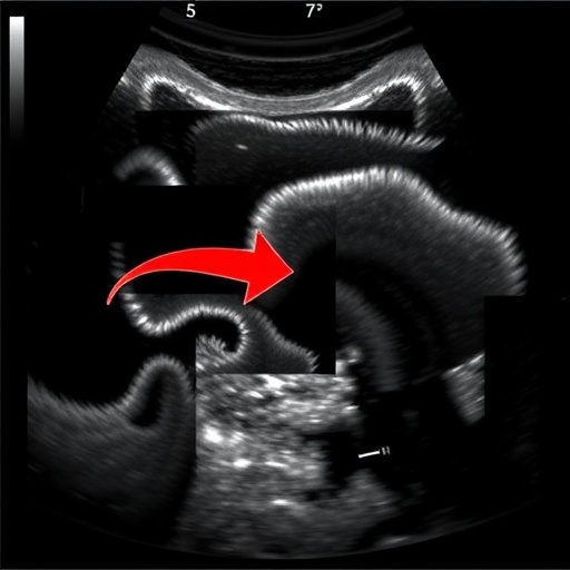
A new study published in Nature Communications is putting artificial intelligence at the center of a difficult medical challenge: determining how severely Schistosoma japonicum has damaged the liver. Led by Xu, Zhang, Wu and colleagues, the research explores deep learning as a tool for precision grading of liver fibrosis from ultrasound images. The work addresses a problem that affects both clinical decision-making and public health surveillance, particularly in regions where schistosomiasis remains a serious disease. Rather than relying exclusively on invasive tissue sampling or broad visual judgments, the approach aims to extract measurable patterns from routine ultrasound examinations and translate them into a more detailed assessment of fibrotic injury.
Schistosomiasis is caused by parasitic flatworms of the genus Schistosoma, not by a virus, and infection can persist for years when exposure continues or treatment is delayed. S. japonicum, found mainly in parts of China and other areas of East and Southeast Asia, can produce chronic inflammation in the liver and the blood vessels surrounding it. The body’s immune response to parasite eggs may lead to the formation of granulomas and progressive scarring. Over time, this process can alter the architecture of hepatic tissue, impair blood flow and contribute to portal hypertension. The severity of fibrosis is therefore more than a descriptive label: it can help clinicians estimate risk, monitor disease progression and select the intensity of follow-up and treatment.
Ultrasound is widely used because it is comparatively affordable, portable and free of ionizing radiation. It can reveal changes in liver texture, surface appearance, vessel structure and the spleen, all of which may be associated with advanced schistosomiasis. Yet ultrasound interpretation is also highly dependent on the experience of the operator. Images may vary according to the machine, examination settings, patient position and the quality of the acoustic window. Early or intermediate stages of fibrosis can be particularly difficult to distinguish because their visual signatures may be subtle and overlap with other forms of chronic liver disease. These limitations create an opportunity for computational systems that can evaluate the full image rather than relying only on a small number of features recognized by the human eye.
Deep learning is a branch of machine learning built around artificial neural networks with many processing layers. In medical imaging, these networks are trained using examples in which an image is paired with a known clinical interpretation or reference diagnosis. During training, the model repeatedly adjusts internal parameters so that its predictions become closer to the assigned labels. Once trained, it can analyze new images and estimate the probability that they belong to particular disease categories. In a fibrosis-grading application, the objective is not simply to identify whether disease is present. The more demanding task is to separate several levels of severity, preserving clinically meaningful distinctions between mild, moderate and advanced structural damage.
The study’s focus on “precision grading” is significant because classification into only two groups can conceal important biological differences. A patient with early fibrosis and another with extensive scarring might both be categorized as having liver involvement, even though their risks and monitoring needs are not identical. A finely calibrated model could potentially support earlier intervention, identify patients requiring specialist assessment and provide a more consistent way to follow changes over time. It might also help reduce variation between hospitals or between sonographers with different levels of experience. However, a model’s usefulness depends on whether its predictions remain reliable across different populations, ultrasound systems and disease patterns, rather than only on its performance within the dataset used to develop it.
Medical-image AI systems commonly rely on convolutional neural networks or related architectures capable of detecting spatial patterns. These networks can learn edges, textures, geometric relationships and larger anatomical arrangements as information passes through successive layers. More recent systems may combine image features with clinical variables, such as age, laboratory measurements or disease history, although the available citation does not specify the architecture used by Xu, Zhang, Wu and their colleagues. Whatever the precise design, the central challenge is to ensure that the network learns signs of fibrosis rather than irrelevant clues. If all severe cases were scanned on one machine, for example, an algorithm might accidentally learn the machine’s imaging signature instead of the underlying pathology.
For that reason, validation is as important as model construction. A convincing clinical AI study generally requires careful separation of training, validation and test data, with the final evaluation performed on images the algorithm has never seen. Researchers may assess accuracy, sensitivity, specificity and agreement with expert or pathological grading. For multiple fibrosis stages, confusion matrices and class-specific performance can show whether the system mainly struggles with neighboring stages or makes larger errors. Calibration is also important: when a model reports a probability, that probability should correspond reasonably well to the actual frequency of disease. External validation at independent centers is especially valuable because it tests whether an algorithm can travel beyond the environment in which it was created.
The reference provided for this research does not include the study’s numerical results, sample size, imaging protocol or details about clinical deployment, so those aspects cannot be responsibly inferred from the citation alone. The publication nevertheless points to a broader transformation in diagnostic medicine. Algorithms are increasingly being developed not to replace clinicians, but to act as decision-support systems that highlight patterns, standardize measurements and identify examinations that deserve closer review. In settings where experienced radiologists or hepatology specialists are scarce, such tools could eventually extend expertise to community clinics. Their role would still require human oversight, transparent reporting and clear safeguards against overreliance on automated scores.
The potential impact is particularly relevant for neglected and geographically distributed diseases. Schistosomiasis control programs often operate across rural areas where advanced imaging, pathology services and specialist care may be limited. A dependable ultrasound-based grading system could make disease assessment more scalable if it can function on commonly available equipment and withstand differences in image quality. It could also support epidemiological studies by making fibrosis grading more reproducible across large groups. Yet implementation would require more than a high-performing algorithm. Health systems would need training, quality control, secure data handling, maintenance and protocols explaining what clinicians should do when the AI’s prediction conflicts with the clinical picture.
The study by Xu, Zhang, Wu and colleagues arrives as researchers continue to test whether artificial intelligence can convert ordinary medical images into richer measures of disease biology. Its subject is a specific parasitic disease, but the underlying question is universal: can machines help clinicians recognize gradual tissue damage before it becomes irreversible, and can they do so consistently enough to improve care? If deep learning can reliably distinguish stages of S. japonicum-associated liver fibrosis, it could strengthen diagnosis, follow-up and resource allocation in affected communities. The next test will be independent clinical validation and real-world evaluation, where accuracy must be matched by explainability, fairness and measurable benefits for patients.
Cite Scienmag News
Blake Davidson. (August 21, 2026). Deep Learning Precisely Grades Schistosoma japonicum Liver Fibrosis Using Ultrasound Images. Scienmag. https://scienmag.com/deep-learning-precisely-grades-schistosoma-japonicum-liver-fibrosis-using-ultrasound-images/
Blake Davidson. "Deep Learning Precisely Grades Schistosoma japonicum Liver Fibrosis Using Ultrasound Images." Scienmag, 21 August 2026, https://scienmag.com/deep-learning-precisely-grades-schistosoma-japonicum-liver-fibrosis-using-ultrasound-images/. Accessed 4 September 2026.
Blake Davidson. "Deep Learning Precisely Grades Schistosoma japonicum Liver Fibrosis Using Ultrasound Images." Scienmag. August 21, 2026. https://scienmag.com/deep-learning-precisely-grades-schistosoma-japonicum-liver-fibrosis-using-ultrasound-images/


