


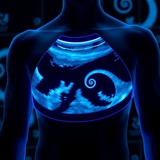
A soft wearable ultrasound patch combined with artificial intelligence could offer intensive-care doctors a faster and safer way to monitor central venous pressure without inserting a catheter into a major vein, according to a new study published in Cyborg and Bionic Systems. The system continuously images blood vessels in the neck, automatically analyzes their changing shape, and estimates whether a patient’s central venous pressure, or CVP, has reached a clinically important level. In a prospective multicenter study of 349 intensive-care patients, the technology achieved an area under the receiver operating characteristic curve of 0.91 in its internal evaluation and 0.87 in an external test, suggesting that it could become a useful screening tool for elevated venous pressure at the bedside.
Central venous pressure reflects the pressure in the right atrium and is widely used as an indicator of venous return, cardiac function, and a patient’s response to fluids or vasoactive medications. The conventional reference method requires placement of a central venous catheter, an invasive procedure that can cause bleeding, infection, thrombosis, pneumothorax, or other complications. Although catheterization remains essential for many critically ill patients, it may be unsuitable or technically difficult in people with coagulopathy, infection, distorted anatomy, or limited venous access. Noninvasive alternatives based on physical examination or intermittent ultrasound can provide valuable information, but they are often operator-dependent and do not deliver uninterrupted monitoring. The new system is designed to bridge that gap by combining a neck-worn imaging device with automated interpretation.
At the center of the platform is an ultra-thin, 128-element linear-array ultrasound transducer developed to sit comfortably over the right side of the neck. The probe operates at a center frequency of 8.5 megahertz, a range that provides a balance between fine spatial resolution and sufficient penetration to visualize the internal jugular vein and common carotid artery. These vessels are important because the internal jugular vein connects directly to the right atrium without intervening valves. Changes in its cross-sectional area can therefore reflect variations in right-sided filling pressure and central venous pressure. The adjacent common carotid artery provides an anatomical reference that helps normalize measurements for differences in probe placement, body habitus, and neck geometry.
The patch’s engineering is intended to preserve image quality during prolonged use. Its acoustic structure includes a dual-layer matching system and a customized backing layer, producing an 85% fractional bandwidth and high sensitivity across a broad range of frequencies. A solid hydrogel coupling material maintains acoustic transmission between the probe and skin without requiring a liquid gel that can dry, leak, or become uncomfortable over time. The electronics and transducer are enclosed in silicone, allowing the device to remain attached while patients move or receive routine care. In feasibility testing, the patch was worn for as long as 24 hours while maintaining stable imaging and acceptable skin comfort, raising the possibility of near-continuous vascular surveillance rather than occasional manual examinations.
Continuous ultrasound, however, generates far more information than a clinician can realistically inspect frame by frame. The device produces cine-loop videos of the jugular vein and carotid artery, and each recording may contain thousands of individual images. To automate this process, the researchers created a semi-supervised artificial-intelligence model called the dual-decoder spatiotemporal attention network, or DSTA-Net. Instead of requiring experts to outline the vessels in every frame, the model uses manual annotations for only about 10% of the images. These key frames correspond to points at which the internal jugular vein is near its maximum or minimum dilation, providing highly informative examples of the vessel’s changing geometry.
DSTA-Net learns from the remaining unlabeled frames through a dual-decoder consistency strategy. A shared encoder first converts each ultrasound image into features that represent vessel boundaries, texture, and surrounding anatomy. Two separate decoders then interpret those features in complementary ways: one incorporates temporal attention to track how structures evolve across consecutive frames, while the other uses a lighter pathway to generate an independent segmentation. The model is trained to make the two pathways agree, allowing unlabeled images to serve as additional learning signals. This approach avoids relying on a continuously updated teacher model, a technique that can be vulnerable to unstable or incorrect predictions in noisy ultrasound data. By exploiting the natural temporal continuity of the cine-loop, the network can follow vessel motion while reducing the annotation burden for medical experts.
Testing indicated that the system could segment the internal jugular vein more accurately than several established deep-learning approaches. On an internal dataset, DSTA-Net achieved a Dice similarity coefficient of 83.5%, while its score on an external dataset was 75.8%. The Dice coefficient measures the overlap between the region identified by an algorithm and the region outlined by an expert, with higher values indicating closer agreement. According to the study, the model improved on the strongest fully supervised baselines, including UNet, Swin-UNet, and DeepLabV3+, by approximately 12 percentage points internally and 9 points externally. It also outperformed semi-supervised systems including UniMatch, DWL, and AllSpark. The model’s derived vascular measurements showed Spearman correlation values above 0.88 for most parameters, and Bland–Altman analyses indicated percentage errors well below the commonly cited 30% threshold for clinical agreement.
The segmented images were converted into five vascular indices: the maximum area of the internal jugular vein, its minimum area, the area of the common carotid artery, the ratio between the maximum jugular and carotid areas, and a jugular-vein area ratio reflecting dynamic changes over time. These measurements were combined with age, body mass index, blood pressure, and heart rate in a second artificial-intelligence system known as a dual-modality multilayer perceptron, or DM-MLP. Unlike image-focused architectures such as convolutional ResNets or vision Transformers, the DM-MLP was designed for structured clinical data. Its Attribute-Mixing operation models relationships among different clinical features, while Case-Mixing refines how each feature is represented across patients. The resulting low-rank architecture uses relatively few parameters while retaining the ability to capture nonlinear interactions, and it outperformed ResNet, DenseNet, and Transformer-based alternatives by roughly 4% to 8% in area under the curve.
The clinical evaluation included 349 intensive-care patients, with 272 enrolled at Shanghai Sixth People’s Hospital and 77 at Shanghai Tenth People’s Hospital. The principal target was elevated CVP, defined as a pressure of at least 8 millimeters of mercury. The model reached an AUC of 0.91 on the internal test set and 0.87 on the external test set, indicating strong discrimination between patients above and below the threshold. Additional analyses using thresholds of 7 and 9 millimeters of mercury produced similarly robust results. SHAP-based interpretability analysis suggested that the ultrasound-derived variables, especially maximum internal jugular vein area and the jugular-to-carotid ratio, contributed more strongly to the predictions than conventional variables such as blood pressure and body mass index. The system processed images at approximately 32 frames per second, or about one frame every 30 milliseconds, on a hospital server, enabling near-real-time analysis.
The researchers stress that the technology is not intended to eliminate central venous catheters in every clinical situation. Instead, it could provide a rapid, repeatable assessment when catheterization is contraindicated, delayed, or unnecessary, and could help identify rising venous pressure before a patient undergoes an invasive procedure. The study remains an early demonstration: its cohort is modest for training modern AI systems, it evaluates diagnostic performance rather than whether the technology improves survival or treatment decisions, and some aspects of the semi-supervised model remain difficult to interpret. Future work will expand the number of participating hospitals, test direct prediction of continuous CVP values rather than categories, add interpretability methods such as Grad-CAM, and assess whether AI-guided monitoring changes fluid and vasopressor management. By uniting wearable ultrasound, temporal image analysis, and clinical prediction, the platform brings automated noninvasive hemodynamic monitoring closer to routine use in acute and critical care.
News Publication Date: August 8, 2026
Web References: DOI: 10.34133/cbsystems.0653
References: Cyborg and Bionic Systems, “AI-Enabled Wearable Ultrasound for Noninvasive Central Venous Pressure Monitoring.”
Cite Scienmag News
Reid Dalton. (August 18, 2026). AI-Powered Wearable Ultrasound Enables Noninvasive Central Venous Pressure Monitoring. Scienmag. https://scienmag.com/ai-powered-wearable-ultrasound-enables-noninvasive-central-venous-pressure-monitoring/
Reid Dalton. "AI-Powered Wearable Ultrasound Enables Noninvasive Central Venous Pressure Monitoring." Scienmag, 18 August 2026, https://scienmag.com/ai-powered-wearable-ultrasound-enables-noninvasive-central-venous-pressure-monitoring/. Accessed 3 September 2026.
Reid Dalton. "AI-Powered Wearable Ultrasound Enables Noninvasive Central Venous Pressure Monitoring." Scienmag. August 18, 2026. https://scienmag.com/ai-powered-wearable-ultrasound-enables-noninvasive-central-venous-pressure-monitoring/


