


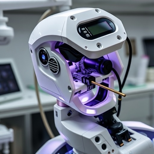
Tooth transplantation has taken a step toward automation after researchers in China demonstrated that an autonomous multi-axis robot can prepare recipient sockets with exceptional geometric precision. In an in vitro study, the robotic system carved nonlinear, surface-conforming osteotomies designed to match the exact three-dimensional shape of a donor tooth. The approach could address one of the most technically demanding stages of tooth autotransplantation: creating a socket that is large enough to receive the tooth, but sufficiently accurate to preserve surrounding bone and protect the periodontal ligament, the living tissue that helps anchor the tooth and support healing.
Tooth autotransplantation involves moving a patient’s own tooth from one location in the mouth to another, often to replace a missing or severely damaged tooth. The treatment is particularly valuable for children and adolescents because it preserves a natural tooth and allows the jaw to continue developing. Unlike dental implants, which may be unsuitable before facial growth is complete, a successfully transplanted tooth can erupt, function, and adapt with the surrounding tissues. Its success depends heavily on minimizing damage to the donor tooth’s root surface and ensuring that the recipient socket mirrors the root anatomy closely enough to permit stable placement without excessive manipulation.
Preparing that socket is difficult because tooth roots rarely have simple, uniform shapes. Single-rooted teeth may still include curved or tapered surfaces, while double-rooted teeth present branching geometries, narrow spaces, and undercuts that are challenging to reproduce with conventional surgical instruments. Dentists generally use a combination of imaging, surgical guides, pilot drilling, and repeated manual refinement. The process relies substantially on operator experience. A socket that is too small can require repeated trial insertions, increasing the time the tooth remains outside the mouth. A socket that is too large can remove unnecessary bone, compromise stability, and create a poor fit that may interfere with periodontal ligament healing.
The research team, led by Dr. Shizhu Bai and Dr. Yimin Zhao of the School of Stomatology at The Fourth Military Medical University in China, designed an autonomous robotic workflow to overcome these limitations. Their findings were published in the International Journal of Oral Science on June 30, 2026. Rather than following a simple straight drilling path, the system executed preplanned nonlinear toolpaths that conformed to the intended surface of the socket. A multi-axis robotic platform controlled the milling instrument’s position and orientation throughout the procedure, allowing it to follow complex root contours and maintain a more consistent relationship with the planned anatomy than a manually refined osteotomy.
The researchers evaluated the system using 40 three-dimensional printed mandibular models representing tooth autotransplantation cases. Half of the models were assigned to the robotic group and half to a static guide-assisted group. Each group included 10 models with single-rooted tooth anatomies and 10 with double-rooted anatomies, allowing the investigators to compare performance in both relatively straightforward and highly complex geometries. Digital planning software was used to position the donor teeth and generate the ideal recipient sockets. To simulate the space needed around the periodontal ligament, the digital donor root surfaces were expanded by 0.5 millimeters, while undercuts were digitally removed to facilitate insertion.
In the conventional workflow, a static guide directed initial pilot drilling, after which the socket was refined freehand. In the robotic workflow, the system translated the digital plan into an automated series of milling movements. After each socket was prepared, the models were digitally scanned and compared with the original surgical plan. The researchers assessed the accuracy of the socket entrance and deepest regions, the angular orientation of the osteotomy, the similarity between planned and manufactured root morphology, the amount of bone removed, and the time required to complete the preparation. This combination of three-dimensional deviation mapping and volumetric analysis enabled the team to measure not only whether the socket began in the correct location, but also whether its deepest and most anatomically important regions matched the intended design.
Both methods achieved similar positional accuracy at the socket entrance, suggesting that static guides can provide reliable initial access. The differences became more pronounced deeper inside the models. Robotic preparation produced significantly smaller deviations at the deepest part of the socket and maintained more accurate drilling angles. The robot-generated cavities also showed stronger volumetric agreement with the planned root shapes, lower surface deviations, and less unnecessary bone removal. In practical terms, the system was better able to reproduce the three-dimensional geometry required for a close anatomical fit instead of creating a generalized cavity that had to be enlarged manually.
The advantage was greatest in double-rooted tooth models, where the branching root structure creates more opportunities for drilling errors and excess bone removal. In these anatomically complex cases, a conventional instrument may not easily reach every surface while maintaining the correct angle, particularly when the socket follows a curved or nonlinear path. The robotic system’s ability to coordinate multiple axes allowed the cutting tool to adjust its orientation continuously as it moved through the planned trajectory. Despite the improved precision, the researchers found that total preparation times were comparable between the robotic and static guide-assisted techniques, indicating that greater accuracy did not come at the cost of a slower laboratory workflow.
The findings suggest that autonomous robotics could make tooth autotransplantation more reproducible and less dependent on individual surgical experience. A socket that closely matches donor-root anatomy may reduce repeated trial insertions, shorten the period during which the tooth is outside the mouth, and preserve more of the bone needed for primary stability. It could also create more favorable conditions for periodontal ligament cells to survive and reorganize after transplantation. However, the results remain a proof of concept rather than evidence of clinical effectiveness. The experiment used printed mandibular models rather than living bone, and the models could not reproduce bleeding, soft-tissue movement, variations in bone density, restricted access, or the biological response of a patient.
Future clinical studies will need to determine whether the geometric improvements observed in the laboratory lead to better healing, greater transplant stability, reduced extra-alveolar time, and improved long-term tooth survival. Researchers will also need to assess how the system performs under real surgical conditions, how easily clinicians can learn to operate it, and whether automated preparation can be integrated into existing digital dentistry workflows without introducing new delays or safety concerns. Even with those questions unanswered, the study points toward a future in which tooth transplantation is planned from high-resolution anatomical data and executed with machine-level consistency. By combining digital imaging, computer-aided design, and autonomous robotic osteotomy, the technology could transform one of dentistry’s most delicate procedures into a more precise, patient-specific, and predictable treatment.
News Publication Date: 30 June 2026
Web References: International Journal of Oral Science article DOI; The Fourth Military Medical University
References: DOI: 10.1038/s41368-026-00446-3
Cite Scienmag News
Ophelia Keating. (August 17, 2026). Scientists Move Closer to Efficient Autonomous Dental Surgery. Scienmag. https://scienmag.com/scientists-move-closer-to-efficient-autonomous-dental-surgery/
Ophelia Keating. "Scientists Move Closer to Efficient Autonomous Dental Surgery." Scienmag, 17 August 2026, https://scienmag.com/scientists-move-closer-to-efficient-autonomous-dental-surgery/. Accessed 3 September 2026.
Ophelia Keating. "Scientists Move Closer to Efficient Autonomous Dental Surgery." Scienmag. August 17, 2026. https://scienmag.com/scientists-move-closer-to-efficient-autonomous-dental-surgery/


