


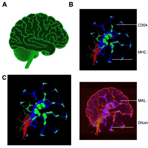
Multiple sclerosis has long been understood as a disease in which immune cells attack the central nervous system, but the cellular conversations that sustain this attack remain incompletely defined. A new study in Nature identifies astrocytes—star-shaped glial cells traditionally associated with neuronal support—as active partners in autoimmune inflammation. The research shows that astrocytes expressing the immune molecules CD40 and major histocompatibility complex class II (MHC-II) can present antigen to CD4+ T cells and intensify pathogenic immune responses in the brain and spinal cord.
The work, led by researchers including J. H. Lee, Z. Li and J. S. Soto, combines several advanced approaches to reconstruct these interactions in experimental autoimmune encephalomyelitis (EAE), a widely used mouse model of multiple sclerosis. The investigators used rabies barcode interaction detection followed by sequencing to identify which cells physically contacted one another. They paired this strategy with single-cell RNA sequencing, in vitro astrocyte–T cell co-cultures and cell-specific CRISPR–Cas9 genetic perturbations performed in vivo. Together, these methods allowed the team to distinguish merely neighboring cells from cells engaged in functional immune communication.
Astrocytes are not professional antigen-presenting cells in the same way as dendritic cells, macrophages or B cells. Under inflammatory conditions, however, they can acquire components of the antigen-presentation machinery. MHC-II molecules display peptide fragments to CD4+ T cells, while CD40 functions as a costimulatory receptor capable of amplifying inflammatory signaling. The study found that astrocytes bearing both CD40 and MHC-II are not passive bystanders in CNS autoimmunity. Instead, they can help activate and maintain T cell responses within nervous tissue, creating a local environment favorable to continued inflammation.
To examine the consequences of direct cell-to-cell contact, the researchers used SorTagging, a system designed to label immune partnerships whenever two cells interact. This enabled them to isolate and analyze CD4+ T cells that had physically contacted astrocytes during EAE. The interacting T cells displayed features associated with pathogenic T helper 17 cells, or Th17 cells. These cells produce inflammatory mediators, including interleukin-17, and are strongly implicated in the development of autoimmune damage in the CNS. Direct contact with astrocytes enhanced the Th17 response, suggesting that astrocytes may help shape the behavior of infiltrating T cells rather than simply responding to signals released by them.
The interaction also changed the astrocytes themselves. The investigators focused on CD40, which is activated when it binds CD40 ligand, or CD40L, expressed by activated CD4+ T cells. According to the study, CD40 stimulation caused astrocytes to accumulate lipid droplets containing the protein PLIN4. Lipid droplets are intracellular organelles increasingly recognized as dynamic metabolic and signaling centers, rather than inert fat stores. In this setting, the droplets appear to supply acetyl-CoA, a central metabolic intermediate that can also serve as a substrate for protein acetylation.
The researchers propose that this metabolic shift strengthens nuclear factor kappa B, or NF-κB, signaling in astrocytes. NF-κB is a major transcriptional regulator of inflammation. Acetyl-CoA generated in association with PLIN4-positive lipid droplets supports acetylation of p65, a key NF-κB subunit. Acetylated p65 can promote inflammatory gene expression and increase the cell’s capacity to present antigen. This creates a potentially self-reinforcing circuit: CD4+ T cells activate astrocytes through CD40L, activated astrocytes accumulate lipid droplets and intensify NF-κB activity, and the resulting antigen presentation further stimulates autoimmune T cells.
To investigate the molecular details of the CD40 pathway, the study combined in vivo subproteomic analyses with AlphaFold-Multimer structural predictions. Subproteomics can reveal changes in defined protein populations and their modifications in specific cellular contexts, while AlphaFold-Multimer can help predict how proteins may assemble or interact. These analyses supported a mechanistic connection between CD40 signaling, PLIN4-positive lipid droplets, acetyl-CoA availability and p65 acetylation. The findings suggest that metabolism is not merely a consequence of astrocyte activation; it may be an essential part of the mechanism that makes these cells immunologically powerful.
Genetic experiments strengthened the causal interpretation. By perturbing genes specifically in astrocytes using CRISPR–Cas9-based approaches, the researchers tested whether the identified pathway influenced disease rather than simply accompanying it. The results indicated that astrocytic CD40 and MHC-II contribute to CNS autoimmunity in EAE. Removing or disrupting components of this system reduced the ability of astrocytes to support pathogenic T cell activity, placing these cells directly within the disease mechanism. The work therefore expands the cellular map of multiple sclerosis beyond infiltrating leukocytes and emphasizes the importance of immune functions acquired by resident CNS cells.
The findings were also examined in human disease material. Single-nucleus RNA sequencing and immunohistochemistry detected astrocytes expressing CD40 and MHC-II, together with lipid-droplet-associated features, in samples from people with multiple sclerosis. Although these observations do not by themselves prove that the same pathway causes disease in patients, they provide evidence that the cellular state identified in mice is relevant to human pathology. The study points to astrocyte–T cell contact, CD40–CD40L signaling and lipid metabolism as possible therapeutic targets, while also highlighting the challenge of interrupting harmful immune activity without compromising the essential support functions of astrocytes. More broadly, the research reveals how a direct conversation between a resident brain cell and an autoimmune T cell can transform local metabolism into a driver of inflammation.
Cite Scienmag News
Juliet Wilcox. (August 5, 2026). CD40+MHC-II+ Astrocytes Present Antigens, Promoting Central Nervous System Autoimmunity. Scienmag. https://scienmag.com/cd40mhc-ii-astrocytes-present-antigens-promoting-central-nervous-system-autoimmunity/
Juliet Wilcox. "CD40+MHC-II+ Astrocytes Present Antigens, Promoting Central Nervous System Autoimmunity." Scienmag, 5 August 2026, https://scienmag.com/cd40mhc-ii-astrocytes-present-antigens-promoting-central-nervous-system-autoimmunity/. Accessed 3 September 2026.
Juliet Wilcox. "CD40+MHC-II+ Astrocytes Present Antigens, Promoting Central Nervous System Autoimmunity." Scienmag. August 5, 2026. https://scienmag.com/cd40mhc-ii-astrocytes-present-antigens-promoting-central-nervous-system-autoimmunity/


