


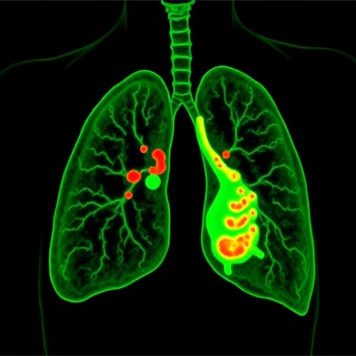
A new study in Nature Communications reports an ultrasensitive way to detect lung inflammation in male mice, using a hybrid imaging strategy that couples immunolabeled magnetic particles with magnetic particle imaging (MPI). The approach is designed to spot inflammatory activity at very low levels, addressing a major challenge for preclinical researchers who need fast, quantitative readouts of disease progression.
The core concept is straightforward but powerful: specially engineered magnetic particles are functionalized with antibodies that recognize inflammatory biomarkers. After administration, the particles accumulate where immune activity is elevated, effectively translating molecular signals from inflamed tissue into a magnetic contrast signal that can be visualized noninvasively.
MPI serves as the readout engine. Unlike conventional imaging modalities that can be limited by background tissue signal, MPI directly detects the magnetic signature of the injected particles. That physical specificity enables high sensitivity and rapid imaging, making it well suited for longitudinal studies in small animals, where repeated measurements are essential.
In the reported work, the researchers focus on lung inflammation in male mice, demonstrating that the immuno-magnetic particles can track inflammatory changes across the respiratory organ. The team emphasizes that the signal correlates with disease-related biological activity, allowing inflammation to be monitored rather than inferred indirectly.
Technically, the study refines both the targeting chemistry and the imaging workflow. Particle–biomarker binding is optimized to ensure selective accumulation in inflamed regions, while MPI acquisition parameters are tuned to maximize contrast and minimize noise. Together, these choices help the system reach detection thresholds that are described as “ultrasensitive.”
Importantly for translational thinking, the method offers a path toward more quantitative immune monitoring. If similar targeting and biocompatibility strategies can be adapted for other biomarkers and disease settings, immuno-MPI could become a versatile tool for testing anti-inflammatory therapies and stratifying disease severity in preclinical models.
The authors also position immuno-MPI as a platform technology. By swapping antibody components, the same imaging framework could be repurposed to interrogate different aspects of inflammation, including distinct immune pathways and stages of disease.
Overall, the results suggest that immuno-magnetic particle imaging can deliver a sensitive, mechanism-linked view of lung inflammation in vivo, potentially accelerating how inflammation is measured in animal studies and how early treatment effects are evaluated in preclinical trials.
Cite Scienmag News
Ophelia Keating. (July 28, 2026). Ultrasensitive Imaging Detects Lung Inflammation in Male Mice With Magnetic Particles. Scienmag. https://scienmag.com/ultrasensitive-imaging-detects-lung-inflammation-in-male-mice-with-magnetic-particles/
Ophelia Keating. "Ultrasensitive Imaging Detects Lung Inflammation in Male Mice With Magnetic Particles." Scienmag, 28 July 2026, https://scienmag.com/ultrasensitive-imaging-detects-lung-inflammation-in-male-mice-with-magnetic-particles/. Accessed 4 September 2026.
Ophelia Keating. "Ultrasensitive Imaging Detects Lung Inflammation in Male Mice With Magnetic Particles." Scienmag. July 28, 2026. https://scienmag.com/ultrasensitive-imaging-detects-lung-inflammation-in-male-mice-with-magnetic-particles/


