


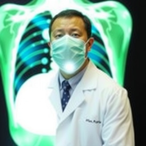
A groundbreaking imaging technology developed by researchers at the University of Oklahoma promises to revolutionize liver transplant evaluations by enabling more comprehensive and rapid assessments of donor organ health. With thousands of patients awaiting liver transplants nationwide, transplant teams urgently need tools that can deliver swift, accurate insights without invasive procedures. The team, led by Dr. Qinggong Tang, has adapted polarization-sensitive optical coherence tomography (PS-OCT)—an advanced imaging modality originally utilized in retinal scans—to noninvasively evaluate donor livers at high resolution.
Traditional liver assessments rely heavily on biopsies, where small tissue samples are extracted and examined for signs of disease or damage. While considered the gold standard, biopsies are inherently limited, providing only a microscopic snapshot of a vast organ that may vary in condition. This narrow sampling can cause uncertainty, sometimes leading to the discard of viable organs. The OU team sought to overcome these limitations by employing PS-OCT, which not only captures detailed cross-sectional images of liver tissue but also measures its polarization properties, revealing subtle structural and biochemical features linked to liver health.
The engineers designed a portable PS-OCT system optimized for clinical and operating room environments, allowing transplant teams to scan liver surfaces quickly and noninvasively. The imaging data are then analyzed using advanced artificial intelligence algorithms, co-developed with Dr. Chongle Pan’s computer science lab. These AI tools quantify critical parameters indicative of hepatic steatosis (fat accumulation) and inflammation, two primary factors influencing liver viability. By training their models on datasets from over thirty clinical cases—comparing PS-OCT results with biopsy outcomes and post-transplant organ function—the researchers demonstrated strong correlations between imaging biomarkers and actual clinical metrics.
Beyond providing a more holistic view of donor liver condition, PS-OCT could enhance decision-making speed, potentially expanding the pool of organs deemed suitable for transplantation by identifying healthy tissue regions missed by biopsy. Such improvements may reduce organ discard rates and improve transplant success. Encouraged by initial findings, the OU team plans a large multisite clinical trial involving institutions like UT Southwestern and the Cleveland Clinic to validate and refine this approach across diverse patient populations.
This work exemplifies OU’s interdisciplinary innovation platform that integrates optical bioimaging, machine learning, pathology, and organ perfusion technology to translate engineering advances directly into clinical practice. The researchers also intend to extend PS-OCT evaluation methods to other organs, including kidneys and lungs, widening the impact of this technology.
Supported by National Science Foundation grants and institutional partnerships, the team’s efforts mark a significant step toward redefining how transplanted organs are assessed. Dr. Tang emphasizes that this is just the beginning, envisioning a future where noninvasive, AI-enhanced imaging facilitates safer, faster, and more effective organ transplantation worldwide.
Keywords
Organ transplantation, Optical coherence tomography, Polarization-sensitive imaging, Liver viability, Artificial intelligence, Biomedical imaging, Noninvasive diagnostics, Medical innovation
Cite Scienmag News
Ophelia Keating. (July 9, 2026). OU Researchers Enhance Imaging Technology for Better Donor Liver Assessment. Scienmag. https://scienmag.com/ou-researchers-enhance-imaging-technology-for-better-donor-liver-assessment/
Ophelia Keating. "OU Researchers Enhance Imaging Technology for Better Donor Liver Assessment." Scienmag, 9 July 2026, https://scienmag.com/ou-researchers-enhance-imaging-technology-for-better-donor-liver-assessment/. Accessed 5 September 2026.
Ophelia Keating. "OU Researchers Enhance Imaging Technology for Better Donor Liver Assessment." Scienmag. July 9, 2026. https://scienmag.com/ou-researchers-enhance-imaging-technology-for-better-donor-liver-assessment/


