Marjolin ulcer is a rare yet formidable form of skin cancer, deceptively slow in its development but aggressive in its behavior once it manifests. Typically arising from chronic wounds, unresolved inflammation, or decades-old burn scars, this malignancy predominantly presents as squamous cell carcinoma (SCC). What sets Marjolin ulcer apart from typical skin cancers is not just its origin but its alarming propensity for local recurrence and metastasis. Medical science continues to grapple with its elusive dormancy and sudden aggressive emergence, making early diagnosis and intervention critical.
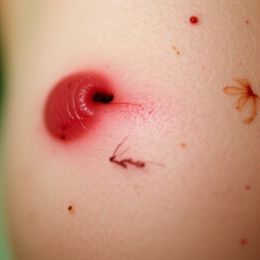
A recent case report sheds light on this insidious pathology through the story of a 72-year-old man presenting with a peculiar lesion on the dorsal aspect of his left thumb. The patient’s lesion evolved insidiously from a seemingly innocuous ulcer, centered in a three-decade-old burn scar. Initially misdiagnosed as a simple infection and treated with topical antibiotics, the lesion’s failure to heal belied an underlying malignant process. Over time, the ulcer expanded, developing a thick, crusted, wart-like appearance measuring nearly three centimeters in diameter, heralding malignancy.
Histopathological examination following biopsy revealed a well-differentiated invasive squamous cell carcinoma, characteristic of Marjolin ulcer. The defining histologic hallmark of such SCCs includes keratin pearl formation, a concentric layering of keratinized cells that signifies squamous differentiation. Under hematoxylin-eosin staining at 100× magnification, these keratin pearls are unmistakable indicators of the tumor’s aggressive phenotype. Despite the invasive carcinoma’s aggressive histopathology, imaging modalities such as magnetic resonance imaging (MRI) and ultrasound confirmed the absence of deeper tissue infiltration or regional lymph node involvement.
This confinement of the malignancy to the superficial layers permitted a surgical approach with curative potential. The patient underwent complete excision of the lesion with clear histologic margins ensuring the removal of all neoplastic tissue. Skin grafting was employed to reconstruct the defect post-excision, facilitating both functional and aesthetic restoration. One year following surgery, the patient remained free of local recurrence or distant metastasis, a positive prognostic indicator for this aggressive tumor type.
Marjolin ulcer is notorious for its unpredictable clinical behavior, with literature citing local recurrence rates ranging from 10% to 37% and distant metastasis in up to 22% of cases—comparatively higher than standard SCCs. This heightened risk profile can be attributed to factors including chronic scar inflammation, delayed diagnosis due to its dormant nature, and the aggressive biological behavior of tumor cells arising from altered scar tissue microenvironments. Recurrence often afflicts the same anatomical site, underscoring the necessity for extended postoperative surveillance.
The molecular underpinnings of Marjolin ulcer, while still under investigation, are believed to involve chronic inflammation-induced genetic mutations. Persistent inflammatory stimuli within scar tissue may promote dysplastic changes in keratinocytes, ultimately driving malignant transformation. Altered expression of tumor suppressor genes and oncogenes has been postulated, contributing to the tumor’s invasive potential and resistance to conventional therapies.
From a clinical perspective, vigilance is paramount. Any new growth, persistent ulceration, or atypical change within longstanding scars warrants immediate clinical assessment and histological evaluation. Early biopsy can unmask otherwise silent malignant transformations before regional or systemic dissemination occurs. The role of advanced imaging cannot be overstated, aiding in delineating tumor extent and guiding surgical planning.
Therapeutic strategies are primarily surgical, emphasizing complete excision with clear margins to reduce recurrence risk. In scenarios where the tumor invades deeper structures or lymph nodes, adjunctive treatments including radiotherapy or chemotherapy may be considered. However, the relative rarity of Marjolin ulcer complicates the establishment of standardized treatment protocols, necessitating individualized and multidisciplinary approaches for optimal outcomes.
The psychological and functional impact on patients with Marjolin ulcers, especially involving the hands or other functionally critical areas, is profound. Surgical removal and subsequent reconstruction require a balance between oncologic control and preservation of limb function, challenging reconstructive surgeons to innovate and adapt. Rehabilitation and long-term follow-up remain integral components of comprehensive patient care.
The described case exemplifies the clinical journey from unnoticed chronic scar to aggressive malignancy, emphasizing the critical need for heightened awareness among both clinicians and patients. In the context of an aging population and rising incidences of chronic wounds and burns globally, the significance of recognizing and managing Marjolin ulcer cannot be overstated. This report serves as a timely reminder that vigilance and early intervention remain the cornerstones of survival against this stealthy adversary.
In conclusion, Marjolin ulcer represents a unique intersection of chronic dermatologic insult and oncologic transformation. It challenges conventional paradigms with its silent latency and sudden, aggressive emergence. Advancements in molecular diagnostics and imaging, combined with meticulous surgical techniques, herald improved prognoses. Yet, the disease’s rarity and complexity necessitate ongoing research and heightened clinical suspicion to effectively combat its morbidity and mortality.
Article Title: Marjolin Ulcer in the Hand
News Publication Date: 29-May-2026
DOI: 10.2738/SKIN.2026.0014
Image Credits: HIGHER EDUCATION PRESS
Keywords: Cell biology, Squamous cell carcinoma, Marjolin ulcer, Skin cancer, Chronic wounds, Burn scars, Keratin pearl, Histopathology
