In a groundbreaking advancement poised to revolutionize interventional radiology, researchers at Washington University in St. Louis have unveiled a portable, point-of-care positron emission tomography (PET) system capable of delivering real-time, high-resolution molecular imaging directly at the patient’s bedside. This cutting-edge technology, recently presented at the Society of Nuclear Medicine and Molecular Imaging (SNMMI) 2026 Annual Meeting, promises to transform clinical workflows by bringing precise molecular imaging to traditionally constrained environments such as intensive care units and operating rooms, thereby enabling more accurate and efficient diagnostic and therapeutic procedures.
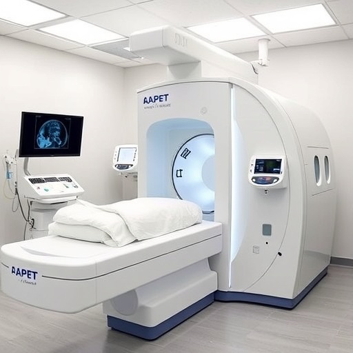
Unlike conventional PET/CT systems, which are large, immobile, and cost-prohibitive for many healthcare institutions, this novel portable PET prototype integrates a benchtop design accompanied by a robotic arm that facilitates flexible positioning of detector panels around the patient. By allowing detector panels to maneuver to arbitrary locations, the system can image virtually any organ of interest with enhanced patient accessibility. This innovative mechanical architecture addresses longstanding challenges of integrating molecular imaging into real-time interventional radiology, where procedure durations and imaging accuracy critically impact clinical outcomes.
Current interventional techniques predominantly rely on anatomical imaging methods such as ultrasound, X-ray fluoroscopy, and computed tomography (CT) for guidance. While these modalities provide structural information vital for navigation, they lack the metabolic and functional insights that molecular imaging offers. Previous studies have demonstrated that dedicated PET/CT-guided interventions yield improved diagnostic precision and treatment accuracy; however, their deployment remains limited due to logistical and financial barriers. The introduction of a portable, cost-effective PET technology capable of bedside application promises to democratize access to molecular imaging and reshape the management of complex clinical cases.
Central to the new system’s performance is a sophisticated imaging workflow that supports interactive PET scanning combined with real-time image reconstruction. During experimental phantom studies, involving radiotracer-filled rod clusters representing heterogeneous tissue structures, the PET detector panels were sequentially positioned at six user-defined locations. Image reconstruction employed an incremental ordered-subsets expectation maximization (OSEM) algorithm, beginning with five iterations using initial data, followed by single iteration updates as new positional data were acquired. This advanced approach leverages the disparity between the longer data acquisition time and much shorter reconstruction phases to continuously refine and display images, offering immediate visual feedback during scanning.
Comparative analyses reveal that the image quality and structural delineation achieved by this real-time, incremental reconstruction framework are on par with conventional full-data maximum likelihood expectation maximization (MLEM) reconstructions, which are performed post-acquisition. Notably, phantom features became distinguishable after data from just three to four detector positions, suggesting the potential for abbreviated scan times without compromising diagnostic integrity. This capability introduces unprecedented flexibility, enabling adaptive scanning protocols tailored to specific clinical needs, reducing patient exposure, and optimizing procedure efficiency.
The scientific team highlights that this interactive scanning methodology fosters a paradigm shift, empowering clinicians with dynamic imaging tools that support decision-making during interventions. The portable PET system’s ability to promptly update molecular images as data accrues promotes a more engaged and responsive clinical workflow, enhancing precision in targeting lesions for biopsies, tumor ablations, or other minimally invasive treatments. Moreover, these advancements pave the way for novel molecular imaging applications that require rapid, bedside assessment without interrupting procedural continuity.
Currently, the research has focused on validating the system using a benchtop prototype; however, efforts are underway to engineer a fully integrated device optimized for human use. Clinical translation is anticipated to begin with initial human imaging studies slated for 2027. The ongoing development phases will address patient safety, ergonomics, and regulatory requirements essential to ensuring efficacy and widespread adoption. By bridging the gap between molecular imaging innovation and clinical accessibility, this technology is set to dramatically improve patient-centered care in interventional radiology.
The implications of bringing real-time PET imaging directly to the bedside extend beyond procedural enhancements. Hospitals constrained by space and resource limitations stand to benefit immensely through improved operational throughput and reduced reliance on centralized imaging suites. This decentralized approach aligns with broader healthcare goals of delivering precision diagnostics in diverse care settings, accelerating treatment timelines, and ultimately improving clinical outcomes. Furthermore, the reduction in logistical burdens may incentivize wider use of molecular imaging, fostering a new standard of care in oncology and other specialties.
Technically, the system integrates advances in detector design, robotic mobility, and algorithmic image reconstruction to overcome previous limitations associated with portable nuclear imaging devices. High sensitivity and spatial resolution are maintained through optimized detector panels, while the robotic arm enables versatile positioning without compromising patient comfort or safety. The real-time reconstruction algorithms are computationally efficient, capitalizing on incremental data inputs to facilitate seamless image updates. The synergy of these components results in a PET device that is both practical and powerful, capable of delivering clinical-grade images in a fraction of the time required by traditional systems.
A key innovation lies in the interactive control over scan positions, which affords operators the ability to customize imaging fields dynamically in response to evolving clinical scenarios. This level of adaptability is unprecedented in PET technology and opens avenues for rapidly tailoring protocols to complex patient anatomy or procedural progress. The system’s robust reconstruction framework ensures that image quality improves progressively as more data is collected, granting clinicians the freedom to conclude scans once sufficient diagnostic confidence is attained, thus enhancing patient experience.
In summary, the development of this portable, robotic-arm-enabled point-of-care PET system marks a significant milestone in molecular imaging technology. By delivering high-quality, real-time molecular images at the bedside with flexible detector positioning and efficient reconstruction algorithms, the system overcomes the historical barriers of size, cost, and accessibility characteristic of conventional PET/CT scanners. This innovation offers the potential to enhance the precision and efficacy of interventional radiology procedures, facilitate bedside diagnostics in space-limited environments, and catalyze new research frontiers in clinical molecular imaging.
The research team at Washington University in St. Louis, led by Dr. Yuan-Chuan Tai and graduate researcher Xiyan Li, emphasizes the transformative clinical impact of this technology, which promises to democratize access to molecular imaging and improve therapeutic outcomes. As prototype development continues towards human applications, the broader medical community eagerly anticipates the integration of this technology into routine clinical practice, heralding a new era of personalized, image-guided care.
—
Subject of Research: Portable point-of-care positron emission tomography (PET) imaging system with real-time image reconstruction.
Article Title: Interactive PET Scanning and Real-Time Image Reconstruction for a Portable Point-of-Care PET System.
News Publication Date: Not explicitly stated; research presented at SNMMI 2026 Annual Meeting.
Web References:
https://www.xcdsystem.com/snmmi/program/UtDKfSi/index.cfm?pgid=3058&sid=53905&mobileappid=5390500000
https://www.snmmi.org
References:
Abstract 262595. “Interactive PET Scanning and Real-Time Image Reconstruction for a Portable Point-of-Care PET System,” Xiyan Li, Samarth Aggarwal, Ling Cai, Pinhuang Wang, Richard Laforest, Joseph A. O’Sullivan, Yuan-Chuan Tai, Washington University in St. Louis.
Image Credits: Courtesy of SNMMI.
Keywords: Positron emission tomography, molecular imaging, medical imaging, point-of-care imaging, interventional radiology, real-time image reconstruction, portable medical devices, robotic imaging systems, image-guided therapy, incremental OSEM reconstruction, adaptive imaging workflows, benchtop PET prototype.
