
Friday, August 28, 2026
Science
No Result
View All Result
  • Login
  • HOME
  • SCIENCE NEWS
  • CONTACT US
  • HOME
  • SCIENCE NEWS
  • CONTACT US
No Result
View All Result
Scienmag
No Result
View All Result
Home Science News Cancer

Nanoscale Nuclear Organization Revealed by High-Resolution Imaging

April 24, 2026
in Cancer
Reading Time: 3 mins read
0
Nanoscale Nuclear Organization Revealed by High-Resolution Imaging
66
SHARES
601
VIEWS
Share on FacebookShare on Twitter
ADVERTISEMENT

In a groundbreaking leap for cellular biology and microscopy, researchers at the Indian Institute of Science (IISc) have developed an enhanced DNA-PAINT imaging technique to visualize multiple biomolecules simultaneously within the nucleus of cancer cells at unprecedented resolution. This advancement opens a new frontier in understanding cellular nuclear architecture by unveiling the spatial organization of critical proteins involved in transcription machinery and nuclear structural integrity, providing one of the most detailed molecular maps of nuclear organization to date.

Cells, the building blocks of life, contain a complex milieu of biomolecules such as proteins and nucleic acids, orchestrating essential biological functions. Traditional microscopy techniques, limited by optical diffraction, have allowed imaging only a handful of molecular species simultaneously, typically two or three. This constraint has restricted researchers’ capability to untangle the intricate interactions and spatial arrangements of molecules essential for genomic regulation and cellular health.

Addressing this challenge, the IISc team employed DNA-PAINT (Points Accumulation for Imaging in Nanoscale Topography), a super-resolution microscopy technique that transcends conventional limits by exploiting the transient binding of fluorescently tagged DNA oligonucleotides to complementary DNA sequences tethered to biomolecules of interest. This method generates blinking fluorescent signals, allowing the precise localization of individual molecules with nanoscale accuracy.

What sets this research apart is the creation of an expanded repertoire of DNA tags, enabling the simultaneous targeting of up to 12 distinct biomolecular species within intact cells. The team engineered five of these probes to possess enhanced binding kinetics — binding faster and remaining attached longer — which significantly improves image quality and spatial resolution down to 3-5 nanometers. Such fidelity in imaging allows researchers to distinguish molecular arrangements at near-molecular scales, previously unattainable by optical methods.

Importantly, the strongly binding DNA tags reduce photodamage by requiring lower laser intensities during imaging, thus preserving the integrity of both the fluorescent probes and the delicate cellular structures. Photobleaching and molecular disruption, common pitfalls in fluorescence microscopy, are minimized, enabling prolonged imaging sessions without compromising data quality.

In addition to improving tagging chemistry, the IISc researchers optimized imaging protocols to accelerate data acquisition. Early implementations of DNA-PAINT required many hours to image a single molecular species due to the need for extensive sampling to achieve high localization precision. The enhanced technique now facilitates rapid, multiplexed imaging of nine different nuclear targets within a timeframe of less than four hours, marking a substantial improvement in experimental throughput.

This high-speed, multi-target imaging capability empowers scientists to observe dynamic cellular processes with outstanding molecular detail. For instance, the team utilized their platform to visualize how cancer cells reorganize their nuclear proteins upon transcriptional inhibition, shedding light on the proteomic rearrangements that underpin cellular response to environmental and pharmacological perturbations.

By generating precise, nanoscale maps of biomolecular distributions in diseased states, this approach offers promising avenues for early disease detection. Subtle alterations in protein interactions and localization, which precede visible symptoms, could serve as biomarkers for cancer and other pathologies, transforming diagnostic paradigms toward earlier and more targeted intervention.

Furthermore, this methodology provides unprecedented insights into the interplay between diverse molecular players within the nucleus. Understanding these intricate relationships is crucial for elucidating mechanisms of gene regulation, chromatin architecture, and the maintenance of genomic stability under physiological and pathological conditions.

The integration of sequence-specific DNA tags with advanced microscopy techniques represents a powerful toolbox for cell biologists and biomedical researchers. It heralds a new era in spatial omics, where the simultaneous imaging of numerous biomolecules at nanoscale resolution paves the way for comprehensive cellular maps, enabling better understanding of complex biological systems in health and disease.

This pioneering study not only enhances the technical capabilities of super-resolution microscopy but also exemplifies the impact of interdisciplinary innovation, where chemistry, molecular biology, and optical physics converge to unravel the complexities of life at the molecular level.

As science continues to push boundaries, technologies like the enhanced DNA-PAINT developed at IISc promise to revolutionize cellular imaging, allowing researchers to peer deeper into the microscopic world and decode the molecular language of cells with unmatched clarity and speed.

Subject of Research: Multiplexed super-resolution imaging of nuclear proteins in cancer cells

Article Title: Nanoscale Nuclear Organization Revealed by High-Resolution Imaging

Article References: Original research article

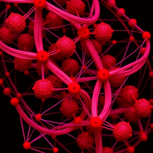
Image Credits: AI Generated

DOI: Not provided

Keywords: advanced cellular microscopy methods, cancer cell biomolecule visualization, cellular nuclear architecture, cellular nuclear integrity analysis, DNA-PAINT fluorescence microscopy, DNA-PAINT technique advancements, fluorescent DNA oligonucleotide imaging, genomic regulation at nanoscale, genomic regulation imaging, high-resolution DNA-PAINT imaging, molecular mapping of nuclear structure, molecular mapping of nucleus, multiplex biomolecule imaging, multiplex biomolecule visualization, nanoscale nuclear organization, nuclear architecture in cellular biology, nuclear structural protein localization, spatial mapping of transcription proteins, super-resolution microscopy in cancer cells, super-resolution microscopy techniques, transcription machinery localization, transcription machinery spatial mapping

Cite this news

SCIENMAG. (April 24, 2026). Nanoscale Nuclear Organization Revealed by High-Resolution Imaging. https://scienmag.com/nanoscale-nuclear-organization-revealed-by-high-resolution-imaging/

SCIENMAG. "Nanoscale Nuclear Organization Revealed by High-Resolution Imaging." Scienmag, 24 April 2026, https://scienmag.com/nanoscale-nuclear-organization-revealed-by-high-resolution-imaging/. Accessed 28 August 2026.

SCIENMAG. "Nanoscale Nuclear Organization Revealed by High-Resolution Imaging." Scienmag. April 24, 2026. https://scienmag.com/nanoscale-nuclear-organization-revealed-by-high-resolution-imaging/


Tags: advanced cellular microscopy methodscancer cell biomolecule visualizationcellular nuclear architecturecellular nuclear integrity analysisDNA-PAINT fluorescence microscopyDNA-PAINT technique advancementsfluorescent DNA oligonucleotide imaginggenomic regulation at nanoscalegenomic regulation imaginghigh-resolution DNA-PAINT imagingmolecular mapping of nuclear structuremolecular mapping of nucleusmultiplex biomolecule imagingmultiplex biomolecule visualizationnanoscale nuclear organizationnuclear architecture in cellular biologynuclear structural protein localizationspatial mapping of transcription proteinssuper-resolution microscopy in cancer cellssuper-resolution microscopy techniquestranscription machinery localizationtranscription machinery spatial mapping
Share26Tweet17
Previous Post

Unveiling Non-Classical Nucleation in Intrinsically Disordered Proteins: From Oligomers and Clusters to Dense Phases

Next Post

Yōni.Fit® Bladder Support for Stress Urinary Incontinence Expands Indication to Include Menstrual Health

Related Posts

Decoy Oligonucleotides in Cancer Therapy: Mechanisms, Challenges, and Clinical Potential
Cancer

Decoy Oligonucleotides in Cancer Therapy: Mechanisms, Challenges, and Clinical Potential

August 27, 2026
Deep learning enables free-breathing MRI, reducing sedation and scan time in children
Cancer

Deep learning enables free-breathing MRI, reducing sedation and scan time in children

August 27, 2026
Commentary examines AI links between lung nodule CT features and cancer-driving genes
Cancer

Commentary examines AI links between lung nodule CT features and cancer-driving genes

August 27, 2026
Indolent γδ Double-Negative T-Large Granular Lymphocytic Leukemia Linked to STAT5B T628S Mutation
Cancer

Indolent γδ Double-Negative T-Large Granular Lymphocytic Leukemia Linked to STAT5B T628S Mutation

August 27, 2026
Cloud Platform Deciphers Tumor Microenvironments and Genomic Landscapes Across Dimensions
Cancer

Cloud Platform Deciphers Tumor Microenvironments and Genomic Landscapes Across Dimensions

August 27, 2026
Mapping NFYA 3′UTRs reveals targetable alternative polyadenylation vulnerability in prostate cancer
Cancer

Mapping NFYA 3′UTRs reveals targetable alternative polyadenylation vulnerability in prostate cancer

August 27, 2026
Next Post
Yōni.Fit® Bladder Support for Stress Urinary Incontinence Expands Indication to Include Menstrual Health

Yōni.Fit® Bladder Support for Stress Urinary Incontinence Expands Indication to Include Menstrual Health

  • Mothers who receive childcare support from maternal grandparents show more

    Mothers who receive childcare support from maternal grandparents show more parental warmth, finds NTU Singapore study

    27656 shares
    Share 11059 Tweet 6912
  • University of Seville Breaks 120-Year-Old Mystery, Revises a Key Einstein Concept

    1061 shares
    Share 424 Tweet 265
  • Bee body mass, pathogens and local climate influence heat tolerance

    682 shares
    Share 273 Tweet 171
  • Researchers record first-ever images and data of a shark experiencing a boat strike

    546 shares
    Share 218 Tweet 137
  • Groundbreaking Clinical Trial Reveals Lubiprostone Enhances Kidney Function

    531 shares
    Share 212 Tweet 133
Science

Embark on a thrilling journey of discovery with Scienmag.com—your ultimate source for cutting-edge breakthroughs. Immerse yourself in a world where curiosity knows no limits and tomorrow’s possibilities become today’s reality!

RECENT NEWS

  • Autodissemination stations curb Aedes notoscriptus and Buruli ulcer risk in urban Australia
  • Label-Free Ovarian Cancer Diagnosis Enhanced by Two-Photon Autofluorescence and Joint Image Processing
  • Programmable Light-Driven Cyclization of Acyclic 1,5-Dienes Creates Rigid Bicyclic Structures
  • Physics-Based Model Predicts Velocity- and Acceleration-Dependent Damping in Adjustable Shock Absorbers

Categories

  • Agriculture
  • Anthropology
  • Archaeology
  • Athmospheric
  • Biology
  • Biotechnology
  • Blog
  • Bussines
  • Cancer
  • Chemistry
  • Climate
  • Earth Science
  • Editorial Policy
  • Marine
  • Mathematics
  • Medicine
  • Pediatry
  • Policy
  • Psychology & Psychiatry
  • Science Education
  • Social Science
  • Space
  • Technology and Engineering

Subscribe to Blog via Email

Enter your email address to subscribe to this blog and receive notifications of new posts by email.

Join 5,150 other subscribers

© 2025 Scienmag - Science Magazine

Welcome Back!

Login to your account below

Forgotten Password?

Retrieve your password

Please enter your username or email address to reset your password.

Log In
No Result
View All Result
  • HOME
  • SCIENCE NEWS
  • CONTACT US

© 2025 Scienmag - Science Magazine

Discover more from Science

Subscribe now to keep reading and get access to the full archive.

Continue reading