Groundbreaking Advancement in Regenerative Medicine: Fully Functional Lab-Grown Esophagus Successfully Implanted in Large Animal Model
In a revolutionary stride for regenerative medicine, scientists at Great Ormond Street Hospital (GOSH) and University College London (UCL) have engineered the first fully functional, lab-grown esophagus capable of seamlessly replacing entire sections of the organ in a large, growing animal model. This advancement marks an unprecedented achievement in tissue engineering and transplantation, demonstrating safe integration and restoration of full physiological function—including swallowing—without the requirement for immunosuppressant drugs.
The esophagus, a vital conduit for transporting food from the mouth to the stomach, plays a critical role in nutrition and growth, especially in pediatric patients. Congenital anomalies such as long-gap esophageal atresia (LGOA), where a significant portion of the esophagus is absent or non-functional, present life-threatening challenges with limited and highly invasive surgical options. The current standard involves complex procedures that often reposition stomach or intestinal tissue to bridge the esophageal gap, bearing the risk of substantial complications, including respiratory issues, gastrointestinal dysfunction, and even uncertain long-term cancer risks.
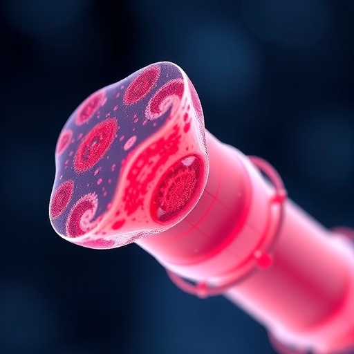
This pioneering research leverages the remarkable biocompatibility and structural similarity between pig and human esophageal tissues to generate customizable, autologous grafts. The critical innovation lies in a multi-step process that begins with decellularization—meticulous removal of all cellular material from donor pig esophageal tissue—leaving behind a sturdy extracellular matrix scaffold that preserves the three-dimensional architecture necessary for organ functionality.
Following scaffold preparation, recipient cells are harvested via a minimally invasive biopsy from the individual requiring surgery. Muscle progenitor cells are isolated and expanded in vitro to achieve quantities sufficient for tissue repopulation. The recipient’s own cells are then seeded onto the decellularized scaffold. This process transforms the tissue into an autologous graft, effectively eliminating immune rejection risks that typically plague transplantation procedures.
To nurture the graft prior to implantation, researchers employed a state-of-the-art bioreactor system. This device perfuses essential growth factors and nutrients through the seeded scaffold, facilitating cellular engraftment, proliferation, and differentiation within a controlled environment. Over approximately one week within the bioreactor, recipient cells proliferate and integrate with the matrix, adopting functional properties essential for muscle contraction, nerve formation, and vascularization.
Implanted into eight growing pigs, these engineered esophagi demonstrated remarkable adaptability and function. Animals recovered well, showing restoration of normal swallowing mechanics and nutritive growth patterns within three months. Notably, the engineered tissue exhibited coordinated muscular contractions and peristaltic movements comparable to those seen in native esophageal tissue. Vascularization and nerve integration were confirmed, underscoring the graft’s capability to sustain itself biologically and functionally in vivo.
One of the breakthrough elements of this study was the application of spatial transcriptomics—a cutting-edge gene-mapping technology that allowed scientists to visualize gene expression patterns within the engineered tissue. This revealed gene activation profiles mirroring those of natural esophageal tissue, verifying not only structural but also molecular fidelity in the regenerated organ.
For pediatric patients, especially newborns and infants with LGOA, this technology offers an unprecedented prospect: a personalized, growing esophageal replacement created from their own cells, capable of integrating seamlessly with their physiology and obviating the need for lifelong immunosuppression. The correlation between the production timeline of these grafts and current treatment protocols suggests clinical feasibility for timely intervention.
Currently, the team is focused on refining this technique—extending graft length, standardizing fabrication protocols, and scaling production while minimizing manual intervention to facilitate clinical translation. Ensuring optimal blood supply functionalization and longitudinal safety remain priorities as preparations are underway for first-in-human trials anticipated within five years.
The impact on affected families could be profound. Traditional surgical solutions impose a heavy burden due to repeated operations and ongoing complications. For children like Casey McIntyre, whose esophageal defect resulted in numerous invasive surgeries and prolonged feeding tube dependence, this technology represents a beacon of hope. A single, effective transplant operation could restore normal feeding and dramatically improve quality of life.
This advancement situates itself at the frontier of regenerative medicine in ways reminiscent of the widespread adoption of porcine heart valves for cardiac patients. By harnessing decellularized xenogeneic scaffolds repopulated with patient-derived cells, the research team at UCL and GOSH is spearheading a transformative approach to organ replacement that may extend to numerous other diseases beyond esophageal malformations.
The study, published in the prestigious journal Nature Biotechnology, underscores the potential of engineering personalized, biocompatible tissues that can grow and function in tandem with the host organism. With ongoing support from GOSH Charity, Oak Foundation, LifeArc, and the Francis Crick Institute, this research embodies a promising future where regenerative therapies provide definitive cures for previously insurmountable pediatric conditions.
Subject of Research: Engineered autologous esophagus transplantation in a large animal model
Article Title: Functional integration of an autologous engineered oesophagus in a large‑animal model
News Publication Date: 20 March 2026
Web References: https://doi.org/10.1038/s41587-026-03043-1
References: “Functional integration of an autologous engineered oesophagus in a large‑animal model,” Nature Biotechnology, 2026
Image Credits: Great Ormond Street Hospital and University College London
Keywords
Regenerative medicine, tissue engineering, esophageal atresia, long-gap esophageal atresia, autologous graft, decellularization, bioreactor, spatial transcriptomics, xenogeneic scaffold, pediatric surgery, organ transplantation, bioengineering
