A Groundbreaking Study Reveals Restorative Brain Network Changes in Neonates with Congenital Heart Disease Following Cardiovascular Surgery
Congenital heart disease (CHD) remains one of the most prevalent birth defects worldwide, representing a significant risk factor not only for cardiac complications but also for disruptions in neurodevelopment. In a pioneering study published in the highly regarded journal JNeurosci, researchers have provided compelling evidence that brain network abnormalities observed in newborns with CHD before surgery show substantial normalization following corrective cardiovascular procedures. This finding not only underscores the profound impact of cardiac anomalies on early brain function but also opens innovative avenues for improving neurodevelopmental outcomes through precise medical intervention.
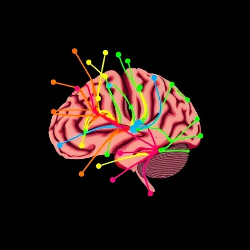
The study, led collaboratively by Jung-Hoon Kim and Catherine Limperopoulos of Children’s National Hospital, capitalized on advanced functional magnetic resonance imaging (fMRI) to explore the resting-state functional brain networks in neonates diagnosed with severe CHD. By systematically comparing these infants’ brain connectivity patterns both prior to and after open-heart surgery with a robust dataset derived from healthy newborn controls, the researchers delineated significant perturbations in key neural circuits underlying sensory processing, motor coordination, and social cognition in affected infants.
Before surgical intervention, resting-state fMRI scans revealed two major functional networks were markedly disrupted in neonates with CHD. These alterations manifest as atypical connectivity patterns that diverge substantially from normative developmental trajectories observed in healthy infants. Specifically, networks crucial for integrating sensory inputs and coordinating voluntary motor activities exhibited diminished connectivity strength and altered spatial organization, indicative of impaired neurophysiological integration during a critical period of brain maturation.
The investigation further highlighted an intriguing asymmetry in brain hemispheric functional organization in preoperative CHD neonates, suggesting a possible lateralized vulnerability to hypoxic or hemodynamic stress inherent to their cardiac conditions. This asymmetry contributes to the complex neurodevelopmental phenotypes frequently observed in this population, including delays in motor skills acquisition and deficits in social behavioral capacities that emerge later in infancy and childhood.
Upon assessing the postoperative conditions several weeks after corrective cardiac surgery, the research team documented a remarkable reversal of these functional abnormalities. The spatial configuration and connectivity strength of the previously disrupted resting-state networks exhibited a significant convergence toward those observed in age-matched healthy controls. Notably, the pronounced hemispheric asymmetry observed preoperatively was substantially reduced, signaling a restoration of more balanced interhemispheric communication pathways.
These neuroimaging findings appear to reflect the brain’s adaptive plasticity in response to improved cerebral oxygenation and cerebral perfusion following surgical correction of the heart defect. The enhanced oxygen delivery reinstates more typical metabolic conditions essential for the maturation and functional integration of neural circuits during the neonatal period — a critical window for brain development.
Limperopoulos emphasizes that the initial connectivity differences noted before surgery likely stem from chronic hypoxia and altered cerebral blood flow patterns associated with cardiac malformation. The restoration of normative brain network structures post-surgery parallels the clinical observation that early corrective interventions can mitigate some of the neurodevelopmental risks in children with CHD, a profoundly hopeful prospect for pediatric cardiac and neurodevelopmental care.
Importantly, Kim notes that leveraging resting-state fMRI as a non-invasive biomarker provides powerful insights into the functional health of the neonatal brain in the context of CHD. Identifying which neural networks are most vulnerable to hypoxic-ischemic injury could lead to earlier and more personalized therapeutic strategies aimed at neuroprotection, rehabilitation, and developmental support.
From a methodological standpoint, the study employed sophisticated analytical techniques to capture subtle alterations in brain connectivity that conventional imaging methods often miss. This approach successfully disentangled complex network interactions and unveiled latent patterns of dysfunction and recovery, thus setting a new standard for neuroimaging research in congenital cardiac disease.
This research also raises critical considerations about the timing and decision-making processes surrounding neonatal cardiac surgery. Limperopoulos advocates for the integration of brain-based biomarkers in clinical protocols to optimize surgical timing, potentially maximizing the neuroprotective benefits of intervention before irreversible neural damage occurs.
Moreover, the identification of neonates who do not exhibit expected postoperative brain network restoration invites avenues for targeted early intervention programs. Such programs could be specifically tailored to reinforce disrupted neural pathways or compensate for persistent deficits, improving long-term neurodevelopmental trajectories and quality of life for these vulnerable children.
Functionally, the study elucidates key developmental neuroscience principles by demonstrating the cerebrum’s sensitivity to systemic physiological states in the nascent stages of life. It highlights the intricate interplay between cardiovascular function and neural system organization, reaffirming the necessity of interdisciplinary approaches that encompass cardiology, neonatology, neurology, and developmental neuroscience.
Collectively, this landmark study not only deepens our comprehension of how congenital heart defects impact neonatal brain organization but also challenges the medical community to incorporate brain connectivity assessments into routine clinical evaluation. Doing so may revolutionize the standard of care by enabling precision medicine strategies that holistically address both cardiac and neurodevelopmental challenges faced by infants with CHD.
As this field advances, further research is warranted to explore the long-term neurodevelopmental outcomes associated with these early brain network changes. Longitudinal studies tracking cognitive, motor, and behavioral functions into childhood and beyond will be essential in validating resting-state connectivity patterns as predictive biomarkers and in guiding therapeutic protocols to foster optimal brain health in this population.
Subject of Research: People
Article Title: Atypical Development of Functional Brain Networks in Neonates with Congenital Heart Disease
News Publication Date: 9-Mar-2026
Web References: http://dx.doi.org/10.1523/JNEUROSCI.1846-25.2026
Image Credits: Kim et al., 2026
Keywords: Congenital heart disease, Neonatology, Cardiovascular disorders, Brain development, Developmental neuroscience
