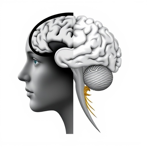
In a remarkable development that promises to reshape our understanding of severe psychiatric disorders, recent research corrections published in Translational Psychiatry illuminate the intricate alterations occurring in the brain’s white matter during early psychosis and schizophrenia. This new insight unfolds against the backdrop of decades of neuroscience investigations emphasizing the critical role of white matter in neural connectivity and cognitive function. White matter, composed primarily of myelinated axons, forms the communication highways of the brain, enabling rapid signal transmission across distant cortical regions essential for integrated brain functioning. The corrected study delves deeply into microstructural changes within this vital neural infrastructure, advancing the narrative beyond traditional gray matter-centric views of psychotic disorders.
Psychosis and schizophrenia have long been framed as disorders characterized by profound disruptions in thought processes, perception, and behavior. Traditional approaches focused largely on neurochemical imbalances and gray matter abnormalities such as cortical thinning or volumetric decreases. However, the vital role of white matter integrity in facilitating efficient neural communication has drawn increasing scientific scrutiny. The corrected findings employed cutting-edge neuroimaging techniques like diffusion tensor imaging (DTI) to map subtle microstructural deviations that may precede or coincide with the onset of psychotic symptoms, offering unprecedented detail about white matter architecture in affected individuals.
The essence of this research correction centers on identifying specific patterns of white matter deterioration during the early stages of psychosis, as well as in fully developed schizophrenia. The findings underscore that altered white matter microstructure is not merely a downstream consequence of disease progression, but a potential biomarker indicating vulnerability to psychosis. Such distinctions are crucial, as they pave the way for earlier diagnostic interventions and open therapeutic windows before irreversible neural damage ensues. The corrected data refine our understanding of which white matter tracts demonstrate the most consistent changes, sharpening the focus on targeted brain networks rather than broad nonspecific deterioration.
Among the most affected white matter tracts are those involved in frontotemporal connectivity. The uncinate fasciculus, which connects the frontal lobe with the temporal lobe including critical limbic structures involved in emotion and memory, exhibits pronounced microstructural alterations. These disruptions align with hallmark symptoms of schizophrenia such as cognitive disorganization, emotional dysregulation, and impaired memory recall. The study correction highlights the importance of preserving these conduits for therapeutic strategies aimed at restoring functional connectivity and mitigating symptom severity, potentially through neuroprotective agents or novel neuromodulation techniques.
Moreover, the corpus callosum—the largest white matter bundle bridging the left and right cerebral hemispheres—shows notable changes in diffusion metrics indicative of compromised integrity. This finding suggests a failure in interhemispheric communication that may underlie the fragmented thought patterns and sensory processing anomalies commonly observed in schizophrenic patients. Importantly, these microstructural changes appear early in the disease course, supporting theories that connect disrupted interhemispheric signaling with the emergence of clinical symptoms in prodromal phases.
From a methodological perspective, this correction emphasizes the significance of rigorous data validation and neuroimaging protocol refinement. The authors employed high-angular resolution diffusion imaging (HARDI) alongside advanced modeling techniques to overcome limitations inherent in standard DTI, such as crossing fiber ambiguities. This methodological enhancement allowed for more precise characterization of white matter microarchitecture, mapping subtle demyelination and axonal damage patterns that were previously obscured. The correction’s transparency in data recalibration further highlights the evolving nature of neuroimaging science and its impact on psychiatric disorder research.
In addition to structural imaging, the correction references emerging multimodal imaging approaches that integrate functional connectivity assessments and microstructural data, offering a holistic view of brain network perturbations. Techniques such as resting-state functional MRI paired with diffusion metrics provide a complementary perspective, revealing how white matter alterations translate into dysfunctional neural circuits. This integrative approach could revolutionize diagnosis by linking microstructural deficits with specific cognitive or behavioral phenotypes, thereby tailoring personalized treatment regimes.
Translational implications stemming from the corrected research encompass early detection strategies using white matter biomarkers. Identifying microstructural deviations in at-risk individuals before clinical symptoms fully manifest offers an unprecedented opportunity to intervene preventively. Such interventions could range from pharmacological treatments aimed at myelin repair to cognitive training designed to enhance compensatory pathways. The correction thus propels the mental health field toward precision psychiatry, where biological underpinnings guide clinical decision-making.
The correction also hints at the heterogeneity of white matter changes among psychosis subtypes, suggesting that future research should focus on stratifying patient populations to elucidate differing neurobiological trajectories. Factors such as age of onset, symptomatology, and environmental influences like stress or substance use may modulate white matter pathology. Understanding these nuances is indispensable for crafting targeted therapies and improving prognostic models.
Critically, this research reintegrates the importance of developmental neurobiology. White matter maturation continues well into the third decade of life, coinciding with the typical emergence window of schizophrenia. Aberrations in neurodevelopmental processes such as oligodendrocyte proliferation and myelin sheath formation could underpin the observed microstructural anomalies. Thus, the correction sheds light on how early life neurodevelopmental insults might predispose individuals to psychosis via disturbed white matter formation, reconciling genetic and environmental risk factors within a unifying framework.
Future research directions inspired by this correction should explore the potential reversibility of white matter disruptions. Animal models and emerging human trials investigating remyelination therapies and neurotrophic factors present promising avenues. Furthermore, longitudinal studies tracking white matter changes over illness progression are essential to discern whether early alterations worsen, stabilize, or potentially recover with appropriate treatment. These pursuits will ultimately inform strategies that prioritize not only symptom management but also the restoration of neural integrity.
In the broader context, this correction contributes significantly to de-stigmatizing psychiatric illnesses by framing them as disorders of brain circuitry rather than mere behavioral anomalies. By elucidating tangible biological alterations, it affirms that psychoses have concrete neuroanatomical substrates, deserving of parity in research focus and funding compared to neurological conditions. This shift could enhance public understanding, reduce prejudice, and encourage individuals to seek help earlier.
Education and public health policies stand to benefit as well from integrating white matter biomarkers into screening programs. The development of noninvasive, accessible scanning technologies could facilitate population-level risk assessment, guiding early interventions and resource allocation. Moreover, linking neuroimaging findings with genetic and metabolic data could enrich comprehensive risk profiles, ushering in an era of multidisciplinary precision medicine within psychiatry.
The corrected article also raises important considerations regarding the ethical deployment of neuroimaging biomarkers. Issues around privacy, consent, and potential discrimination based on biological risk necessitate careful governance. Researchers, clinicians, and policymakers must collaborate to establish frameworks ensuring responsible use that maximizes patient benefit while safeguarding individual rights.
Finally, this landmark correction not only refines technical understanding but also revitalizes hope for patients and families grappling with psychosis and schizophrenia. It highlights that the brain’s white matter, once considered a passive background structure, plays a dynamic and pivotal role in psychiatric disease mechanisms. Recognizing this is a critical step toward developing novel, effective treatments that target underlying neural pathologies, promising improved outcomes and quality of life in the future.
Subject of Research: White matter microstructure alterations in early psychosis and schizophrenia.
Article Title: Correction: White matter microstructure alterations in early psychosis and schizophrenia.
Article References:
Pavan, T., Alemán-Gómez, Y., Jenni, R. et al. Correction: White matter microstructure alterations in early psychosis and schizophrenia. Transl Psychiatry 15, 469 (2025). https://doi.org/10.1038/s41398-025-03740-6
Image Credits: AI Generated


