


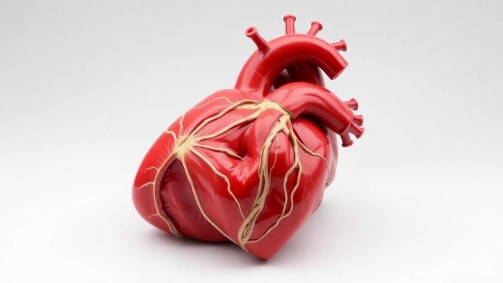
For more than a century, the dissection laboratory has stood as the undisputed centerpiece of medical anatomy education, a rite of passage in which students first confront the messy, wondrous reality of the human body. But a new study from researchers at Drexel University’s College of Medicine suggests that when it comes to actually learning anatomical structure, digital technology may finally be catching up to the cadaver. In a controlled experiment with incoming first-year medical students, a team led by Stephanie A. Waldman and Caitlin A. Howe found that students taught using virtual three-dimensional scans of prosected human hearts performed just as well on a practical anatomy exam as students taught with the physical specimens themselves. The findings, published in the journal Frontiers of Digital Education, arrive at a moment when medical schools worldwide are rethinking how, and how much, anatomy should be taught with digital tools.
The research team’s starting point was a practical one. As medical curricula increasingly incorporate new technologies, educators need evidence about whether those technologies actually help students learn, and whether students themselves would embrace them. To that end, the Drexel group built a database of virtual 3D scanned prosections—cadaveric specimens that have been professionally dissected for teaching purposes and then captured as digital three-dimensional models—for students to use while studying gross anatomy. Prosections are a precious and finite resource: each one requires hours of skilled dissection, and the supply of donor bodies is limited. A high-fidelity digital replica, by contrast, can be viewed by unlimited numbers of students, at any time, from any location, without degrading.
To test whether the digital versions could stand in for the real thing, the researchers recruited twenty-nine incoming first-year medical students before they had begun formal anatomy coursework. The participants were divided into two groups: fifteen students would be taught using physical prosections of the external heart, and fourteen would learn from virtual 3D scans of the same prosections. The study was deliberately structured in four stages, allowing the team to measure learning gains with unusual rigor for an educational technology trial.
First came a pre-test, administered to all participants, that used both physical prosections and images of the virtual 3D scans to probe what the students already knew about external heart anatomy. Then each group received a teaching session using its assigned modality—the physical group handling and studying the actual dissected hearts, the virtual group working with the 3D scanned models. Immediately afterward, all students took a post-test identical to the pre-test, again assessed on both physical specimens and digital images. Finally, every participant completed a survey about their experience and attitudes toward the technology.
The exam results delivered the study’s headline finding: both groups improved dramatically, and neither format held an advantage. Students taught with physical prosections raised their scores to an average of 42.6 percent, with a standard deviation of 17.9, while students taught with the virtual 3D scans reached an average of 44.3 percent, with a standard deviation of 24.0. The difference between the groups was not statistically significant, but the improvement within each group was, with post-test scores rising significantly above pre-test baselines regardless of whether students had prior anatomy experience. In other words, the digital models taught anatomy just as effectively as the cadaveric specimens they replicated—a result that carries real weight given how often virtual resources are dismissed as inferior substitutes.
Perhaps more striking was what students believed about the technology after using it. Those who had learned with the virtual 3D scans were significantly more likely to agree with the statement that they would be able to sufficiently learn anatomy using 3D scans, rating their agreement at 4.1 on a Likert-type scale compared with 3.0 among students taught with physical prosections. First-hand experience with the digital resource, it seems, converted skeptics into believers. Confidence in one’s own learning is not the same as learning itself, but the alignment of subjective confidence with objective performance in the virtual group is exactly the pattern educators hope to see when introducing a new tool.
Yet the survey also revealed a firm boundary to students’ enthusiasm. Regardless of which group they had been assigned to, participants disagreed with the idea that they would have a similar laboratory experience if they learned from 3D scans instead of dissection—the physical group averaging 2.1 and the virtual group 2.5 on the agreement scale. The dissection laboratory, it appears, offers something the students regard as irreplaceable, whether that is the tactile engagement with real tissue, the ritual of working with a donor body, or the collaborative culture of the anatomy lab itself. This finding echoes a long tradition in medical education scholarship that frames dissection not merely as a means of learning structures but as a formative professional experience in its own right.
At the same time, students saw a clear and valuable role for the digital resource alongside traditional teaching. Both groups agreed that they would use the virtual 3D scans to prepare for the dissection laboratory and for practical and written examinations, with ratings of 4.5 in the physical group and 4.9 in the virtual group. Rather than viewing the technology as a replacement, the students positioned it as a complement—a way to preview structures before entering the lab, review them afterward, and study at times when the laboratory is closed and the prosections are locked away. This blended model aligns with a broader trend in anatomy education, where meta-analyses of three-dimensional visualization technologies have generally found modest but real learning benefits when such tools are integrated with, rather than substituted for, conventional instruction.
The context for this study matters. The COVID-19 pandemic forced anatomy courses around the world online almost overnight, accelerating adoption of virtual and augmented reality resources and prompting a wave of research into their effectiveness. Surveys of anatomy education before and during the pandemic documented a rapid, sometimes chaotic shift toward digital delivery, and questions about the appropriate balance between cadaveric and digital learning have remained contentious ever since. Studies of computer-generated three-dimensional models, immersive virtual reality, photogrammetry-based training tools, and web-based interactive visualization have produced a mixed but increasingly encouraging picture, with the Drexel results adding a rare head-to-head comparison using authentic scanned prosections rather than idealized computer renderings.
The study’s authors are careful about what their data can and cannot show. With twenty-nine participants, the sample is small, and the standard deviations—especially the wide spread of 24.0 points in the virtual group—indicate considerable variability in how much individual students gained from each format. The subject matter, the external heart, represents one region of gross anatomy, and results might differ for more spatially complex regions such as the temporal bone or the brachial plexus. The students were also tested immediately after teaching, leaving open questions about long-term retention. Still, the core conclusion is difficult to dismiss: on a practical examination, virtual 3D scans of prosections performed on par with the physical specimens from which they were made.
For medical schools weighing expensive investments in digital anatomy infrastructure, the message is nuanced but actionable. Virtual 3D prosection databases can serve as legitimate learning tools, capable of producing measurable gains in anatomical knowledge equivalent to those achieved with physical specimens, and students who use them come away confident in their value. But the same students who endorse the technology also insist that it should not replace the dissection laboratory itself. The future of anatomy education, if the learners have their say, is not digital versus cadaveric but digital and cadaveric—scanned hearts on the screen before the exam, real ones on the table in the lab, and a generation of physicians trained with the best of both.
Subject of Research: Comparing the effectiveness of virtual 3D scans and physical cadaveric prosections for teaching external heart anatomy to medical students
Article Title: A Practical Examination and Feedback Survey Evaluating Learners Taught Using Physical Prosections vs. 3D Models of Prosections of the External Heart
Article References: Waldman, S. A., Sejdiu, Z., O’Hara, S. M., Shumsky, J. S., & Howe, C. A. (2025). A Practical Examination and Feedback Survey Evaluating Learners Taught Using Physical Prosections vs. 3D Models of Prosections of the External Heart. Frontiers of Digital Education, 2(3), Article 27. https://doi.org/10.1007/s44366-025-0064-9
Image Credits: AI Generated
DOI: 10.1007/s44366-025-0064-9
Keywords: anatomy education, medical education, 3D scanning, prosections, gross anatomy, virtual dissection, heart anatomy, cadaveric specimens, student performance, digital learning, Drexel University, practical examination
Cite Scienmag News
Courtney Benton. (October 2, 2026). 3D Scans Match Cadaver Prosections for Learning Heart Anatomy, Study Finds. Scienmag. https://scienmag.com/3d-scans-match-cadaver-prosections-for-learning-heart-anatomy-study-finds/
Courtney Benton. "3D Scans Match Cadaver Prosections for Learning Heart Anatomy, Study Finds." Scienmag, 2 October 2026, https://scienmag.com/3d-scans-match-cadaver-prosections-for-learning-heart-anatomy-study-finds/. Accessed 4 October 2026.
Courtney Benton. "3D Scans Match Cadaver Prosections for Learning Heart Anatomy, Study Finds." Scienmag. October 2, 2026. https://scienmag.com/3d-scans-match-cadaver-prosections-for-learning-heart-anatomy-study-finds/


