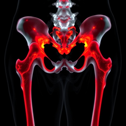
In a groundbreaking study, researchers led by Hao et al. have uncovered the potential of ultrasound radiomics in identifying microstructural changes in the femoral head associated with developmental dysplasia of the hip (DDH). This condition, prevalent among infants and young children, can lead to significant long-term complications if not diagnosed and treated early. The study, published in Pediatric Radiology, highlights the innovative use of advanced ultrasound technologies to detect subtle changes in bone structure that traditional imaging may overlook, thereby paving the way for more effective and timely interventions.
Ultrasound has long been utilized in pediatric imaging owing to its safety profile and absence of ionizing radiation. However, the advent of radiomics—a field that extracts large amounts of quantitative features from medical images—has revolutionized how we analyze ultrasound images. This study leverages radiomic techniques to assess the femoral head’s microstructure in patients afflicted with DDH, aiming to enhance diagnostic accuracy and improve patient management strategies.
The researchers utilized a cohort of pediatric patients diagnosed with DDH, employing high-resolution ultrasound imaging to capture intricate details of the femoral head. Through sophisticated image processing algorithms, they extracted numerous radiomic features from the ultrasound images. These features encapsulate various morphological and texture-related parameters, offering insights into the underlying bone structure that could signify early pathological changes treated with more precision.
One of the pivotal goals of this research was to create a reliable classification model that could distinguish between the normal microstructure of the femoral head and those exhibiting signs of dysplasia. By applying machine learning techniques to the radiomic data, the research team was able to train predictive models demonstrating high accuracy. This step represents a significant advance over standard diagnostic tools, providing clinicians with an advanced mechanism to evaluate the presence and severity of DDH.
Moreover, the significance of identifying microstructural changes cannot be understated. Early detection of these alterations through non-invasive ultrasound techniques can facilitate timely interventions, potentially averting the complications associated with untreated developmental dysplasia. Traditional imaging methods often fail to reveal critical early signs, resulting in delayed diagnoses that can lead to painful surgeries and extended recovery periods for young patients.
The researchers meticulously analyzed the ultrasound images, focusing on key areas of interest within the femoral head. Their findings indicated that specific radiomic features showed a strong correlation with established indicators of dysplasia, thus validating the potential of ultrasound radiomics as a complementary, diagnostic tool. The advancement of machine learning algorithms has enabled improved processing capabilities, allowing for a more nuanced interpretation of the radiomic data.
In the evolving landscape of pediatric radiology, the implications of Hao et al.’s research extend beyond merely enhancing diagnostic accuracy. The study sets a precedent for integrating artificial intelligence into clinical workflows, providing promising avenues for future research in ultrasound radiomics. For instance, further exploration could reveal how these techniques can be scaled to other pediatric musculoskeletal conditions, offering a broader application of this innovative approach.
As the field continues to evolve, the role of collaboration between radiologists, orthopedic surgeons, and data scientists will become increasingly vital. The integration of their insights can lead to better-designed studies that comprehensively address the challenges in detecting developmental dysplasia and implementing effective treatment protocols. Such interdisciplinary efforts can facilitate the development of an optimal framework for adopting ultrasound radiomics in routine clinical practices.
The study conducted by Hao et al. serves as a validation for the transformative potential of combining traditional imaging with advanced computational techniques. The detectable microstructural changes in the femoral head can lead to preemptive measures, making this research a landmark endeavor in pediatric healthcare. This approach aligns well with a patient-centered healthcare model that prioritizes early detection and personalized treatment plans.
In addition, this research underscores the importance of ongoing education and training for medical professionals who will interpret these complex radiomic data. As the technology advances, so too must the skill set of practitioners who rely on these images to inform their clinical judgments. Familiarity with radiomic features and their clinical significance will increasingly define best practices in musculoskeletal imaging.
Perhaps one of the most promising aspects of this study is its potential to influence the development of standardized protocols for using ultrasound radiomics in pediatrics. A unified framework could help streamline care pathways, making interventions more efficient and effective across varied healthcare settings. Such standardization could also facilitate the sharing of data among institutions, promoting collaborative research efforts that could yield more comprehensive insights into pediatric conditions.
The findings of Hao et al. also raise intriguing questions for future research. Could ultrasound radiomics be adapted for other aspects of pediatric health concerns or even be applied in adult populations? What other conditions could benefit from this analytical approach? How can subsequent studies improve the machine learning models to increase predictive power? Each of these questions presents opportunities for further investigation, signaling a path toward innovation and progress in the field of radiology.
As the world of technology and medicine converges, the importance of integrating artificial intelligence continues to grow. The implications of Hao et al.’s study extend beyond immediate clinical applications; they demonstrate the importance of embracing new technology for better patient outcomes. As fields collide, enriched methodologies will redefine our understanding and management of complex health issues in pediatric populations.
Groundbreaking studies like this one inspire optimism in the realm of pediatric radiology, showcasing the ever-expanding boundary of what is possible. By employing ultrasound radiomics, researchers have uncovered new potentials for better understanding developmental dysplasia of the hip, evidencing a commitment to evolve practices that prioritize patient well-being.
Thus, as we look toward the future, the intersection of imaging, data analysis, and artificial intelligence will undoubtedly shape a new landscape in medical diagnostics that enhances accuracy and augments therapeutic strategies, all while ensuring the ultimate aim of healthcare: improving the quality of life for young patients facing challenges today.
Subject of Research: Ultrasound radiomics for identifying microstructural changes in developmental dysplasia of the hip.
Article Title: Ultrasound radiomics for identifying microstructural changes in the femoral head with developmental dysplasia of the hip.
Article References:
Hao, J., Wang, X., Pan, Z. et al. Ultrasound radiomics for identifying microstructural changes in the femoral head with developmental dysplasia of the hip. Pediatr Radiol (2025). https://doi.org/10.1007/s00247-025-06358-4
Image Credits: AI Generated
DOI: https://doi.org/10.1007/s00247-025-06358-4
Keywords: Pediatric radiology, ultrasound radiomics, developmental dysplasia of the hip, machine learning, bone microstructure.
