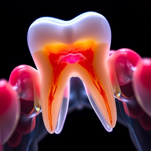
A groundbreaking study led by Dr. Han-Sung Jung at Yonsei University College of Dentistry in South Korea has unveiled remarkable insights into the intricate coordination of tooth development. This research significantly alters our understanding of how teeth, bone, and gums form, emphasizing the critical role of positional information among embryonic cells. Dr. Jung’s team meticulously analyzed how the spatial arrangement of these cells influences their developmental pathways, potentially paving the way for innovative approaches in dental medicine.
Tooth formation is not merely a straightforward process; it is a dynamic and multifaceted journey consisting of various stages, including the bud, cap, and bell phases, followed by root development. Each stage is characterized by complex epithelial-mesenchymal interactions that dictate how the tooth will ultimately form. Researchers have hypothesized for years that the location of a cell within a developing embryo dictates its fate, primarily due to differential signaling molecule concentrations. This study dives deeper into that notion, investigating the molecular pathways that govern cell behavior as teeth begin to shape.
Historically, scientists identified a tooth’s evolution from a small bud formed by outer epithelial cells, which mature into deeper mesenchymal cells. These cells undergo a transformative process, curving to create cap and bell shapes, ultimately leading to a fully formed tooth. Dr. Jung and his team extended this understanding by probing how spatial positioning influences the fate of dental cells, with their findings published in the esteemed International Journal of Oral Science. This research is poised to reshape the way we perceive dental embryology.
The ramifications of this research are profound. Lead author Dr. Jung stated that the team aimed to clarify how positional identity along the lingual-buccal axis could determine the distinct fates of dental mesenchyme. This understanding has broad implications, from developmental biology to clinical applications concerning dental regeneration and repair. By elucidating the intrinsic mechanisms that guide tooth development, the findings provide a foundational understanding that may transform stem-cell therapies and regenerative approaches in dentistry.
The investigative team meticulously separated mesenchymal cells along the lingual and buccal sides during the cap and bell stages of mouse embryonic development. By employing advanced RNA sequencing techniques, they profiled gene expression variances based on spatial positioning and developmental stages. The results revealed stark differences; lingual cells primarily drove tooth formation and structural development, whereas buccal cells focused more on stem cell activity and the support of surrounding tissues.
In an intriguing twist, when the researchers haphazardly mixed cap-stage tagged buccal and lingual cells from genetically engineered mice, the expectation was that they would lose their specialized functions. Contrary to this assumption, the tagged lingual cells managed to localize and reorganize themselves, regenerating the characteristics essential for proper tooth formation. This phenomenon, described as cellular self-organization, indicates a remarkable resilience and adaptability inherent within these cells—a finding that subverts previous assumptions regarding cellular differentiation.
Moreover, the researchers conducted extensive analyses on signaling molecules in each cell population, uncovering profound insights into the biological pathways that guide dental development. WNT signaling and R-spondins (specifically Rspo1/2/4) were significantly more prevalent in lingual cells, who exhibited heightened proliferation rates, reduced apoptosis, and enhanced migration capabilities, all contributing to efficient tooth formation. In stark contrast, buccal cells were discovered to express higher levels of BMP inhibitors, resulting in slower proliferation and migration rates, more aligned with the construction of supportive bone and tissue structures.
Ultimately, the authors proposed a novel model based on the lingual-buccal axis, suggesting that dental cell positioning has critical implications for both tooth and surrounding tissue formation. By drawing attention to the signaling pathways, such as WNT/BMP interactions, governing cellular outcomes, the study invites further exploration into the molecular underpinnings of tooth development. This foundational knowledge may inspire new directions in tissue engineering and regenerative medicine.
As the researchers continue to investigate these cellular dynamics, the insights gained could catalyze advancements in stem cell-based approaches to dental restoration and repair. The promise of harnessing these biological mechanisms could lead to innovative therapies for tooth regeneration, offering hope to patients facing tooth loss or damage. This research marks a significant step forward in dental science, marrying fundamental biological understanding with potential clinical applications.
Continued exploration in this field holds the key to unraveling even further complexities of dental development. As scientists delve into the world of molecular signaling and cellular positioning, the future of dentistry may well evolve into an era where regenerative medicine and cellular therapies become the norm rather than the exception. The ongoing research led by Dr. Jung and his colleagues sets the stage for exciting breakthroughs that could ultimately redefine the landscape of dental health and restoration.
In conclusion, the findings from this study offer a compelling glimpse into the future of dental medicine, signaling that a deeper understanding of embryonic development may lead to innovative solutions in dental care. As research efforts continue to unravel the intricate dance of cellular signaling and positional identity in embryonic development, the possibilities for regenerative dental therapies and their applications become increasingly promising.
Subject of Research: Animal tissue samples and their implications for tooth development.
Article Title: Prespecified dental mesenchymal cells for the making of a tooth.
News Publication Date: 9-Oct-2025.
Web References: Not available yet.
References: DOI: 10.1038/s41368-025-00391-7.
Image Credits: Credit: Thomas Ricker from Flickr.
Keywords
Tooth development, embryonic cells, dental mesenchyme, WNT signaling, regenerative medicine, cellular self-organization, stem cell therapy, molecular pathways, dental restoration, research study, positional identity, tissue engineering.


