In a groundbreaking advance for respiratory medicine, researchers have unveiled a comprehensive spatial single-cell atlas of the human lung that illuminates the intricate regional diversity within both healthy and diseased states. This pioneering work deciphers the cellular architecture with unprecedented resolution, uncovering spatial variations that redefine our understanding of lung biology and pathology. The atlas lays a crucial foundation for developing precision therapies targeting lung diseases such as chronic obstructive pulmonary disease (COPD), asthma, and pulmonary fibrosis, by mapping how individual cells interact across distinct anatomical regions.
Utilizing sophisticated single-cell RNA sequencing techniques coupled with spatial transcriptomics, the study meticulously catalogues cell types and states across different lung regions. Unlike prior bulk analyses that averaged signals from numerous cells, this spatially resolved approach preserves cellular context, allowing researchers to pinpoint exactly where pathological changes occur within the lung’s complex landscape. This level of detail reveals distinct cellular neighborhoods characterized by unique molecular signatures, highlighting previously unrecognized heterogeneity within the tissue microenvironment.
The research team employed cutting-edge multiplex imaging technologies, enabling simultaneous visualization of numerous cell markers across lung tissue slices. This multiplexing, combined with computational modeling, was instrumental in reconstructing a three-dimensional cell atlas that accurately reflects the spatial distribution of diverse cell populations. The resolution achieved bridges an essential gap between histology and molecular biology, providing a dynamic view of cellular interactions governed by spatial proximity and signaling gradients.
One of the key discoveries includes identifying unique cellular compositions linked to different lung regions such as the alveolar space, bronchi, and vasculature. Each area exhibits distinct cell type abundances and gene expression profiles, underscoring a finely tuned regional specialization that supports diverse respiratory functions. Moreover, disease-specific alterations emerge distinctly within these regions, offering insights into how lung pathologies manifest and progress in a spatially confined manner.
In patients affected by chronic lung diseases, the atlas reveals profound shifts in cellular ecosystems. For example, fibrotic lesions display altered stromal and immune cell configurations that differ substantially from healthy counterparts. These localized changes influence tissue remodeling and inflammatory responses, emphasizing the need to consider spatial context when designing therapeutic interventions. The atlas thereby provides a molecular roadmap for targeted drug delivery to affected lung compartments.
Importantly, the study highlights the interplay between epithelial cells and resident immune populations, demonstrating how their spatial proximity modulates immune surveillance and tissue repair. Disruptions in these interactions correlate with pathological inflammation and impaired regeneration, hallmark features in several lung conditions. Understanding these spatial dynamics opens new avenues to modulate immune responses therapeutically for better outcomes.
Advanced bioinformatics tools played a pivotal role in dissecting the complex datasets generated. Machine learning algorithms were applied to classify cell types automatically, predict cell-cell communication networks, and delineate spatial gene expression gradients. This computational prowess enabled the conversion of massive data into accessible, interpretable maps that detail the lung’s intricate cellular geography both in health and disease.
The atlas also surfaces rare cell types and transitional states that were not easily detectable using previous methods. These elusive cell subsets may hold critical roles in maintaining lung homeostasis or orchestrating pathological cascades. Their identification sets the stage for future investigations into their biological functions and potential as therapeutic targets.
Translationally, this spatial atlas is poised to revolutionize diagnostic approaches by linking molecular signatures with histopathological features. By integrating spatially resolved molecular profiling into clinical workflows, physicians could better stratify patients based on regional tissue alterations, predict disease trajectory, and personalize treatments accordingly. This paradigm shift underscores the clinical relevance of high-resolution, single-cell spatial mapping technologies.
Furthermore, the study propels the field of regenerative medicine by pinpointing niches of progenitor and stem-like cells across lung regions. Understanding these spatially distinct cellular reservoirs is critical for devising strategies to enhance lung repair and regeneration following injury or chronic damage. Such knowledge accelerates the quest to develop cell-based therapies tailored to specific lung compartments.
The collaborative effort behind this research brought together experts from molecular biology, computational science, and pulmonary medicine, embodying a model for interdisciplinary innovation. This confluence of expertise ensured rigorous validation and broad applicability of the atlas across diverse populations and disease conditions, paving the way toward equitable healthcare solutions.
Looking forward, the spatial single-cell atlas will serve as a dynamic resource for the scientific community, offering a foundational reference to explore lung biology in finer detail. Continued refinement of spatial transcriptomic methods promises even higher resolution and greater throughput, expanding our capacity to decode respiratory pathophysiology comprehensively.
In summary, this seminal study marks a paradigm shift in lung research by integrating spatial biology with single-cell resolution to reveal the complex and regionally distinct cellular tapestry of the human lung. Its implications resonate beyond the laboratory, steering toward transformative clinical applications that aim to alleviate the global burden of lung diseases through precision medicine rooted in spatial cellular insights.
Subject of Research:
Human lung cellular architecture and its regional variations in health and disease.
Article Title:
Spatial single-cell atlas reveals regional variations in healthy and diseased human lung.
Article References:
Firsova, A.B., Marco Salas, S., Kuemmerle, L.B. et al. Spatial single-cell atlas reveals regional variations in healthy and diseased human lung. Nat Commun 16, 9745 (2025). https://doi.org/10.1038/s41467-025-65704-0
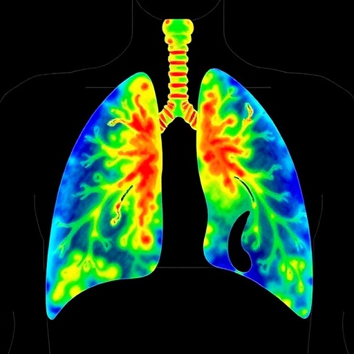
Image Credits: AI Generated
DOI: https://doi.org/10.1038/s41467-025-65704-0
