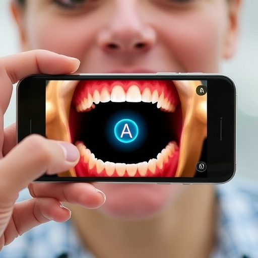
In the realm of cancer detection, oral cancer remains a challenging adversary. Despite the accessibility of the oral cavity for examination, many cases are diagnosed in late stages, when treatment options become limited and survival rates diminish significantly. This alarming scenario is partly due to the difficulty frontline dental professionals face in distinguishing harmless oral lesions from those potentially indicative of malignancy. While dentists and hygienists regularly screen patients, the nuanced differentiation between benign and precancerous conditions often requires specialized expertise that is not always readily available in general dental clinics. Addressing this critical gap, a pioneering team from Rice University, led by Rebecca Richards-Kortum, has developed an innovative, low-cost, smartphone-based imaging platform termed mobile Detection of Oral Cancer, or mDOC, designed to enhance early detection and referral accuracy for suspected oral cancer.
The mDOC system represents a convergence of cutting-edge optical imaging and artificial intelligence. It employs a dual approach utilizing both white light and autofluorescence imaging to capture and analyze oral lesions. Autofluorescence imaging, a technique that illuminates tissue with blue light, reveals characteristic changes in tissue fluorescence emission that occur during abnormal cellular and molecular transformations. Precancerous and cancerous tissues often demonstrate diminished autofluorescence compared to surrounding healthy mucosa, providing a non-invasive window into early disease processes. However, this imaging method alone suffers from specificity limitations; inflammatory and other benign conditions can also produce decreased fluorescence, potentially leading to false alarms. To circumvent these challenges, mDOC integrates a deep learning algorithm trained to interpret imaging data in concert with critical patient-specific risk factors such as age, smoking history, and lesion location. This synergy aims to refine diagnostic precision beyond what imaging or clinical judgment can achieve independently.
A recent study published in Biophotonics Discovery details the rigorous evaluation of the mDOC platform within real-world dental care settings. Researchers enrolled fifty patients across two community dental clinics in Houston, Texas, systematically capturing images from up to five oral sites per individual. Expert clinicians reviewed these images independently, providing referral decisions that served as the gold standard for algorithm training and validation. The study employed a rehearsal training strategy, combining newly acquired patient data with historical datasets from both high-risk and healthy populations. This methodological approach ensured that the model could generalize effectively to community dental practices where the prevalence of suspicious lesions is relatively low, a crucial factor for preventing excessive false positives that could overwhelm clinical workflows.
The performance metrics achieved by the mDOC system underscore its promise and potential impact. On a holdout testing dataset representing a typical low-prevalence clinical environment, the algorithm attained an area under the receiver operating characteristic curve (AUC-ROC) of 0.778, indicating good discriminative ability. Sensitivity reached 60 percent, meaning the system successfully identified the majority of lesions warranting specialist referral as adjudicated by experts. Specificity was even more impressive at 88 percent, reflecting the device’s aptitude for minimizing unnecessary referrals and related patient anxiety or resource burden. Intriguingly, the algorithm’s performance surpassed that of dental providers involved in the study, who exhibited zero sensitivity despite perfect specificity. This finding highlights the difficulty clinicians face in recognizing early oral malignancy without adjunctive tools, and the transformative potential for mDOC to act as a decision-support system.
Though the system showed high accuracy, it was not without imperfections. Among the five referral sites the algorithm identified, two were misclassified but corresponded to lesions that resolved by the time of specialist consultation. This temporal discrepancy raises the possibility that mDOC accurately flagged lesions that would have warranted evaluation but spontaneously regressed, underscoring the complexity of oral lesion dynamics and the challenge of establishing definitive diagnosis at a single time point. Conversely, the device generated 21 false positives, cases where the system suggested referral but expert review did not deem it necessary. These instances indicate room for further algorithmic refinement to enhance specificity and reduce potential over-referral, which remains a critical target for reducing patient burden and optimizing clinical utility.
One of the most compelling advantages of the mDOC platform is its operational efficiency. The average imaging duration of just 3.5 minutes per patient is well-suited to the fast-paced environment of dental clinics, facilitating seamless integration into routine screening without disrupting workflow. This efficiency combined with the portability and accessibility of smartphone hardware enables widespread deployment, including in underserved communities where access to oral cancer specialists is limited or nonexistent. By democratizing early cancer detection, mDOC promises to bridge healthcare disparities that contribute to late diagnosis and poor outcomes in vulnerable populations.
Technically, the image analysis workflow within mDOC is meticulous and robust. Clinically significant anatomic sites are digitally masked, cropped, and resized to standardize input for the deep learning model, ensuring consistent data quality regardless of variability in image acquisition. This preprocessing pipeline allows the network to focus computational resources on diagnostically relevant regions rather than extraneous background information. The deep neural network then synthesizes the processed images with structured clinical metadata, leveraging multimodal data fusion to enhance prediction accuracy. This architectural design reflects a sophisticated understanding of oral lesion pathology and leverages recent advances in artificial intelligence viability for medical imaging applications.
Looking ahead, the research team envisions several avenues for advancing the mDOC system. Incorporating more granular patient history, such as detailed tobacco and alcohol use metrics, HPV status, and genetic predispositions, could further tailor the predictive algorithms and reduce false positive rates. Additionally, continued accumulation of diverse clinical data will support iterative algorithm retraining, improving robustness and adaptability across varied demographic and clinical contexts. Expanding the platform’s capacity to longitudinally monitor lesions could also provide dynamic risk assessments, facilitating personalized surveillance strategies for patients at elevated risk of malignant transformation.
The implications of mDOC’s success extend beyond oral cancer detection. Its conceptual framework—combining smartphone-based optical imaging with advanced machine learning and patient risk profiling—serves as a blueprint for similar strategies targeting other cancers and diseases accessible via non-invasive imaging. Such innovations herald a new era of point-of-care diagnostics characterized by scalability, affordability, and enhanced clinical decision support. The ongoing convergence of photonics, artificial intelligence, and mobile health technology is poised to redefine preventive medicine, empowering non-specialist practitioners with tools previously confined to specialty centers.
In summary, the development and evaluation of the mDOC system mark a significant step forward in oral healthcare innovation. By providing dental professionals with a reliable, easy-to-use tool capable of detecting lesions warranting specialist referral, mDOC addresses a profound unmet need and holds potential to shift the paradigm of oral cancer diagnosis towards earlier, more accurate detection. As oral cancer incidence continues to challenge public health worldwide, technologies like mDOC offer hope for improving survival outcomes through timely intervention, especially in resource-limited settings. This study serves as a compelling demonstration of how interdisciplinary research can create impactful solutions that translate seamlessly into everyday clinical practice.
Subject of Research: Early detection and referral decision support for oral cancer through mobile imaging and machine learning.
Article Title: Optimization of a mobile imaging system to aid in evaluating patients with oral lesions in a dental care setting.
News Publication Date: October 6, 2025.
Web References: https://www.spiedigitallibrary.org/journals/biophotonics-discovery/volume-2/issue-04/042307/Optimization-of-a-mobile-imaging-system-to-aid-in-evaluating/10.1117/1.BIOS.2.4.042307.full
References: R. Mitbander et al., “Optimization of a mobile imaging system to aid in evaluating patients with oral lesions in a dental care setting,” Biophoton. Discovery 2(4), 042307 (2025), doi: 10.1117/1.BIOS.2.4.042307.
Image Credits: R. Mitbander et al., doi 10.1117/1.BIOS.2.4.042307
Keywords
Oral cancer, Smartphones, Imaging, Data analysis, Optics


