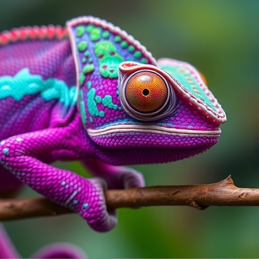
Chameleons have long captivated biologists and naturalists with their extraordinary abilities, particularly the near-360-degree rotation of their eyes and their capacity to independently scan their environment. Despite centuries of study dating back to ancient Greek philosophers, the detailed anatomy that underlies this remarkable ocular mobility has remained elusive — until now. Recent advances in modern imaging techniques have uncovered a groundbreaking discovery: chameleons possess uniquely coiled optic nerves, a morphological trait unseen in other lizards and rare across the animal kingdom.
Juan Daza, an associate professor at Sam Houston State University and lead author of a newly published study, explains that these coiled nerves enable the chameleon’s eyes to swivel independently like surveillance cameras when searching for prey. Once a target is located, the eyes synchronize, directing their focus forward to accurately calibrate the trajectory of the chameleon’s ballistic tongue. This hunting mechanism depends heavily on the precision of ocular coordination, highlighting the functional importance of their specialized optic nerve anatomy.
This discovery was first made in 2017 when Edward Stanley, director of the Florida Museum of Natural History’s digital imaging lab, noticed the distinctive coiled structure in a CT scan of the minute leaf chameleon (Brookesia minima). The optic nerves spiraled in a complex pattern, a configuration that had never been documented in other reptiles. Given the long history of chameleon research, the revelation that such a significant anatomical feature had been overlooked surprised both scientists.
The oversight is partially attributed to limitations of traditional dissection methods. Physical dissection tends to disrupt or damage the fragile optic nerves, preventing clear observation of their true morphological arrangement. However, the advent of computerized tomography (CT) scanning technology allows researchers to non-invasively peer inside the specimens, preserving and revealing inner structures with unprecedented clarity. This imaging modality has revolutionized anatomical studies by providing three-dimensional reconstructions without any physical alteration of the specimen.
The coiled configuration of the optic nerve acts as a biological slack mechanism, permitting extended eye rotation without undue strain or damage to the nerve fibers. Daza analogizes the structure to the spiral cords of early telephone handsets, which provide flexibility and additional length without tangling. Such an adaptation is critical since chameleons possess limited neck mobility, compelling them to rely on eye movement for environmental surveillance rather than head turns, unlike animals such as owls or lemurs.
Intriguingly, comparative analyses of digital CT scans across more than thirty species of lizards and snakes, including various chameleons, confirmed that this coiled optic nerve morphology is exclusive to chameleons. These species showed significantly longer and more intricately convoluted optic nerves than their non-chameleon relatives, confirming that this trait is a distinctive evolutionary innovation within the chameleon clade.
Developmental studies tracing embryonic stages of the veiled chameleon (Chamaeleo calyptratus) reveal that the nerves begin as straight fibers during early embryogenesis but gradually develop coils prior to hatching. This temporal progression indicates that the coiling is a specialized developmental adaptation rather than a static ancestral trait. Hatchlings enter the world equipped with fully mobile, independently rotating eyes, immediately benefiting from this sophisticated neural architecture.
Historical scholarship reveals that, although chameleons have fascinated scientists for millennia, earlier anatomists struggled to characterize their optic nerve anatomy accurately. Aristotle mistakenly thought these reptiles lacked optic nerves, whereas later thinkers such as Roman physician Domenico Panaroli postulated the presence of non-crossing optic nerves to rationalize the eyes’ independent movements. Isaac Newton repeated this hypothesis in his seminal work Optiks, yet even foundational scientists failed to detect the nerve coils discovered today.
The oldest known chameleon fossils date back to the early Miocene epoch, approximately 16-23 million years ago, a period after the development of many arboreal adaptations but too incomplete to resolve the evolutionary origin of the coiled nerve architecture precisely. Although fossilized soft tissues like nerves rarely fossilize, this morphological novelty likely emerged as a functional compensation aligned with the chameleon’s ecological niche—a tree-dwelling lifestyle demanding extreme ocular mobility.
CT scans from initiatives like openVertebrate (oVert), an extensive open-access database fostered by the Florida Museum of Natural History and partners, have democratized access to 3D vertebrate anatomical data, facilitating such discoveries. By leveraging vast repositories of digital specimens, researchers can investigate rare features at scale, revealing evolutionary innovations beyond the reach of conventional anatomy.
The functional implications of coiled optic nerves extend beyond chameleons, prompting broader questions about the interplay of nerve morphology and sensory flexibility in vertebrates. For example, humans possess long optic nerves enabled by the spacing of eye sockets and brain orientation, allowing eye rotations without nerve damage. Rodents, too, exhibit wavy optic nerve fibers that provide similar flexibility. However, the precise spiral coiling seen in chameleons is a unique evolutionary solution to the constraints imposed by limited neck movement.
This breakthrough reshapes our understanding of chameleon neuroanatomy and ocular mechanics, revealing a sophisticated physiological adaptation that supports their niche specialization. As Stanley notes, the fusion of cutting-edge imaging technology with open data initiatives is the key to unraveling longstanding biological mysteries. The scientific community eagerly awaits follow-up research exploring whether convergent evolutionary mechanisms exist in other arboreal reptiles or vertebrates with similar ecological demands.
Despite all the monumental strides in the biological sciences inspired by titanic figures like Aristotle and Newton, nature continues to harbor secrets even in well-studied species. The revelation of coiled optic nerves in chameleons underscores the untapped potential of digital anatomy and integrative techniques to illuminate evolutionary novelties that eluded detection through centuries of traditional approaches.
The authors detailed these findings in the journal Scientific Reports, providing compelling evidence for coiled optic nerves as a uniquely chameleonic trait integral to their ocular mobility and hunting strategy. This discovery not only enriches the field of vertebrate morphology but also exemplifies how technology can propel fundamental biological insights into the limelight, inviting scientists worldwide to re-examine long-held assumptions and explore the complexities hidden within familiar organisms.
Subject of Research: Specialized optic nerve morphology in chameleons
Article Title: A new twist in the evolution of chameleons uncovers an extremely specialized optic nerve morphology
News Publication Date: 10-Nov-2025
Web References:
References: Collins et al., 2025, Scientific Reports
Image Credits: Collins et al., 2025
Keywords: Optics, Science history, Reptiles, Computerized axial tomography, Museums, Biodiversity, Evolution, Fossils
