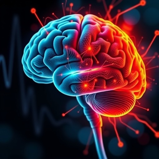
In the intricate landscape of human memory, the ability to suppress fear-related recollections after traumatic or unpleasant experiences is not merely beneficial but essential for maintaining psychological health and adaptive behavior. Without this inhibition, individuals may be predisposed to debilitating psychiatric conditions such as anxiety disorders and depression. While decades of animal research have elucidated that fear extinction involves the formation of new, context-dependent memories that override original fear responses, direct electrophysiological evidence pinpointing such mechanisms in humans has remained elusive—until now.
A landmark study published recently in Nature Human Behaviour by a collaborative team of neuroscientists and psychologists from the Universitat Autònoma de Barcelona and Ruhr-Universität Bochum offers a groundbreaking glimpse into the electrophysiological underpinnings of fear extinction in the human brain. Utilizing advanced neurotechnological methods, the researchers have charted the dynamic neural processes that govern how aversive memories are altered and ultimately suppressed in people, opening promising avenues for the treatment of fear-based psychopathologies.
Central to this novel investigation is the deployment of Representational Similarity Analysis (RSA), a cutting-edge analytical technique that delves deeper than traditional neuroimaging approaches. RSA transcends mere activation maps by enabling scientists to dissect how different brain regions encode and represent specific episodic memories. As Daniel Pacheco-Estefan, the study’s lead author and a cognitive neuroscientist at UAB, elaborates, “RSA allows us to reveal the fine-grained, mechanistic architectures of memory representations, moving beyond general activation to understand how distinct memories are instantiated in neural patterns.”
The experimental design crafted by the team was meticulously intricate, incorporating multiple cues and contextual variations across three pivotal phases: memory acquisition, extinction learning, and subsequent retrieval testing. This multifaceted setup enabled the researchers to replicate the complexities inherent in classical conditioning paradigms within the human brain, and perhaps more importantly, to empirically test hypotheses that were, until now, predominantly validated only in animal models.
Remarkably, the study was conducted with the participation of 49 patients undergoing clinical treatment for epilepsy, who had intracranial electrodes implanted for seizure management. This unique access allowed the researchers to directly record electrical activity from brain structures intimately linked with fear processing—specifically, the amygdala and hippocampus. Participants were exposed to neutral images such as a hairdryer, a fan, and a toaster. During the acquisition phase, some of these images were systematically paired with an aversive auditory stimulus, conditioning a fear response. Crucially, in the extinction phase, the same images were presented without the unpleasant sound, thereby facilitating the process of attenuating the conditioned fear response.
One of the most salient findings involved heightened theta oscillations emerging predominantly in the amygdala during the extinction learning phase when participants were re-exposed to previously fear-associated cues. Theta rhythms, known to play critical roles in emotional memory encoding and retrieval, here seemed to signal a “safety” marker, denoting the suppression of the initial fear response. This electrophysiological signature underscores the amygdala’s pivotal function in not only coding fear but also in signaling the attenuation or extinction of aversive memories.
Furthermore, representational analyses revealed increased similarity between neural responses to items that had been paired with negative stimuli, suggesting that the brain maintains a generalized representational template for unpleasant experiences. This neural convergence potentially elucidates why traumatic memories tend to intrude involuntarily and indiscriminately across diverse contexts in individuals suffering from conditions such as post-traumatic stress disorder (PTSD). Dr. Pacheco-Estefan posits that this represents a neural basis for the persistence and pervasive reactivation of unwanted fear memories.
The study also intricately examined the contextual dependency of extinction memories. During subsequent testing, fear memories were more likely to resurface outside the original extinction context, a phenomenon consistent with the notion that safety memories are “bound” to the specific situational parameters in which extinction learning occurred. As highlighted by Nikolai Axmacher, lead investigator at Ruhr-Universität Bochum, “From the patient’s perspective, the safe context formed during therapy might be perceived as a unique episodic event rather than a broadly applicable experience, which complicates the generalization of extinction learning into everyday life.”
This context specificity carries profound implications for therapeutic strategies that aim to extinguish pathological fear. It explains, at a neural level, why many individuals experience relapse or return of fear symptoms once removed from structured therapeutic environments. Such insights challenge clinicians to develop interventions that can generalize extinction memories beyond the confines of clinical settings, thereby promoting lasting recovery.
Beyond its clinical ramifications, this seminal work invites broader contemplation about the fundamental mechanisms of episodic and autobiographical memory in the human brain. The demonstrated ability to trace representational dynamics electrophysiologically affords researchers an unprecedented window into how complex emotional memories are encoded, maintained, transformed, and extinguished. This biomechanistic framework not only advances cognitive neuroscience but also enriches our understanding of human psychological resilience.
The use of intracranial electrodes in clinical populations represents both a methodological strength and ethical consideration. Access to precise neural recordings from deep brain structures in humans remains rare and ethically constrained; thus, this study’s design stands as a paragon of translational neuroscience that bridges preclinical animal findings with clinical neurobiology in humans. It validates the transferability of earlier animal models and confirms that key elements of fear extinction are conserved across species, albeit with nuanced contextual modulations.
In summation, these pioneering findings illuminate the dynamic interplay between neural oscillations, memory representations, and context dependency during fear extinction in humans. They enrich the theoretical canon of memory research and carry transformative potential for psychiatric treatment. As anxiety and trauma-related disorders continue to exert a substantial global health burden, understanding the exact neural substrates that govern fear remission empowers the development of novel, targeted therapies—potentially harnessing neuromodulation or memory reactivation techniques informed by electrophysiological signatures.
The study thus stands as a remarkable exemplar of how interdisciplinary collaboration, advanced neurotechnologies, and carefully designed experimental paradigms can collectively decode the labyrinthine processes underlying one of the most essential functions of the human brain: the capacity to learn from and ultimately overcome fear.
Subject of Research: People
Article Title: Representational dynamics during extinction of fear memories in the human brain
News Publication Date: 5-Aug-2025
Web References: https://doi.org/10.1038/s41562-025-02268-5
Keywords: Psychological science, Fear
