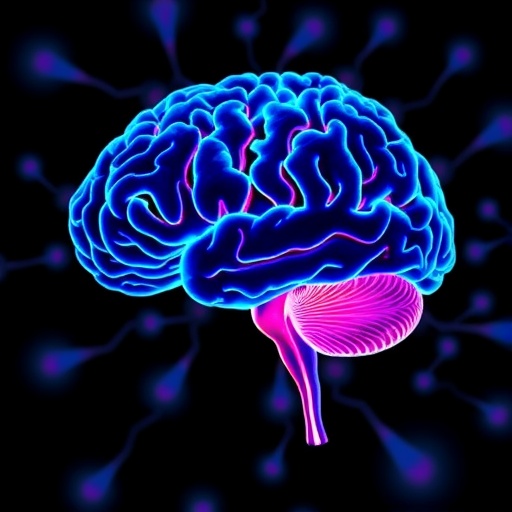
In a groundbreaking collaboration between Karolinska Institutet and Yale University, researchers have unveiled an unprecedented molecular atlas detailing the postnatal development of the mouse brain and its intricate response mechanisms to inflammation. The study, recently published in the prestigious journal Nature, presents a multidimensional perspective that not only charts temporal brain development but also reveals how certain genetic and molecular programs, critical during early neurodevelopment, can be reactivated during neuroinflammatory processes.
Brain development is a marvel of biological precision, encompassing a symphony of cellular differentiation, spatial distribution, and functional maturation. However, capturing these multifaceted processes simultaneously across different molecular layers has been a formidable challenge. Addressing this, the scientific team pioneered a novel methodological approach termed spatial tri-omics. This cutting-edge technique synchronously measures gene expression, epigenetic regulation, and protein synthesis within distinct anatomical brain regions. By harnessing this integrative platform, researchers achieved an unparalleled resolution in mapping the molecular choreography underlying brain maturation and immune response.
Focusing on developmental stages from birth through juvenile periods, the research incorporates comprehensive analyses of both murine and human cerebral tissues. This cross-species approach enhances the translational relevance of findings, offering insights that bridge fundamental neurobiology with human neuropathology. The innovative spatial tri-omics methodology allowed for tracking not just static snapshots but the dynamic evolution of cellular and molecular states during critical developmental windows.
One of the pivotal observations centers on the corpus callosum, a heavily myelinated brain structure facilitating interhemispheric communication. The process of myelination, whereby oligodendrocytes envelop neurons with insulating myelin sheaths, is essential for rapid and efficient nerve signal propagation. However, this vital process is vulnerable in various neurological disorders, notably multiple sclerosis (MS), where autoimmune attacks precipitate demyelination and neurodegeneration. Utilizing a mouse model engineered to disrupt myelination in targeted brain regions, the researchers discerned notable patterns of microglial activation, the resident immune cells of the central nervous system.
Remarkably, microglial activation was not confined to regions of direct injury. The study uncovered that neuroinflammation propagates beyond localized damage sites, implicating a sophisticated and possibly systemic communication network within the brain. This phenomenon challenges existing paradigms that traditionally viewed neuroinflammatory responses as spatially confined. Professor Rong Fan from Yale University highlighted that this discovery underscores a complex interregional signaling architecture that could recalibrate our understanding of brain immune surveillance and response.
Further illuminating this complexity, the research revealed that genetic programs operative during early brain development become re-engaged in the adult brain under inflammatory conditions. This reactivation of developmental molecular pathways suggests that neuroinflammation might co-opt mechanisms originally intended for brain growth and maturation, potentially influencing disease progression or recovery. Professor Gonçalo Castelo-Branco of Karolinska Institutet emphasized that such mechanistic insights could provide new therapeutic avenues, offering strategies to modulate or harness these reactivated pathways to promote remyelination or protect against immune-mediated damage.
The implications of these discoveries are profound for demyelinating diseases like MS. Understanding that inflammation can spread to anatomically distant brain regions may explain the multifocal nature of MS lesions observed in patients. Moreover, the re-engagement of developmental programs during neuroinflammation opens doors to innovative treatments aimed at reprogramming the brain’s intrinsic repair mechanisms. These findings could stimulate the development of biomarkers predictive of disease activity and severity, as well as tailored interventions that target both immune modulation and regenerative processes.
From a methodological standpoint, the spatial tri-omics approach stands as a transformative tool in neuroscience research. By concurrently integrating transcriptomics, epigenomic landscapes, and proteomics with spatial localization, the approach captures the multilayered regulation of neural cells in situ. This comprehensive profiling facilitates a deeper understanding of cell-type-specific responses and intercellular communication in both healthy development and disease states, setting a new benchmark for future studies in complex tissues.
The research team comprised a diverse group of experts spanning neurobiology, immunology, and bioinformatics, highlighting the necessity of interdisciplinary collaboration to tackle such multifaceted biological questions. Co-first authors Di Zhang and Leslie Kirby contributed significantly to the advancement and application of the spatial tri-omics platform, reflecting the critical role of early-career investigators in driving scientific innovation.
This ambitious study received robust funding support from a consortium of international agencies, including the Swedish Research Council, the Swedish Brain Foundation, the Knut and Alice Wallenberg Foundation, the European Union’s Horizon Europe programme, and the U.S. National Institutes of Health. Transparency regarding potential conflicts of interest was maintained, with disclosures noting Professor Castelo-Branco’s shares in Nexus Epigenomics and Professor Fan’s advisory roles in biotechnology firms.
In summary, this landmark investigation not only charts the dynamic molecular landscape of brain development but also elucidates how neuroinflammatory insults can reactivate dormant developmental programs and propagate across brain regions. These revelations hold transformative potential for understanding and eventually mitigating the pathological processes that underlie devastating neurological diseases like multiple sclerosis. The integration of advanced spatial multi-omics techniques with classical neurobiology represents a powerful paradigm shift in the quest to unravel the brain’s complexity.
Subject of Research: Brain development and neuroinflammation dynamics
Article Title: Spatial dynamics of brain development and neuroinflammation
News Publication Date: 5-Nov-2025
Web References:
https://www.nature.com/articles/s41586-025-09663-y
References:
Zhang, D., Rubio Rodríguez-Kirby, L. A., Lin, Y., Wang, W., Song, M., Wang, L., Wang, L., Kanatani, S., Jimenez-Beristain, T., Dang, Y., Zhong, M., Kukanja, P., Bao, S., Wang, S., Chen, X. L., Gao, F., Wang, D., Xu, H., Ma, C., Lou, X., Liu, Y., Chen, J., Sestan, N., Uhlén, P., Kriegstein, A., Zhao, H., Castelo-Branco, G., & Fan, R. (2025). Spatial dynamics of brain development and neuroinflammation. Nature. https://doi.org/10.1038/s41586-025-09663-y
Keywords:
Brain development, Neuroinflammation, Spatial tri-omics, Multiple sclerosis, Microglia activation, Myelination, Oligodendrocytes, Epigenetic regulation, Transcriptomics, Proteomics, Nervous system, Demyelinating diseases
