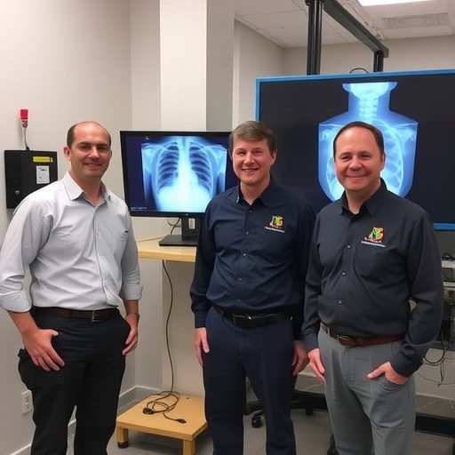
In the late 19th century, the scientific world was forever altered by the discovery of X-rays, a revolutionary tool for imaging and diagnostics. This breakthrough, introduced by German physicist Wilhelm Röntgen, unveiled a new frontier in both medicine and research. Yet, as technology has evolved, the fundamental principles of X-ray generation have remained relatively unchanged, leaving room for innovation. Researchers at Sandia National Laboratories, led by optical engineer Edward Jimenez, have now introduced a pioneering technology that has the potential to redefine X-ray imaging — Colorized Hyperspectral X-ray Imaging with Multi-Metal Targets (CHXI-MMT).
Within the scope of this groundbreaking research lies the intricate interplay between various metals and the distinct colors of X-ray light they emit. This innovative method aims to transition X-ray imaging from its traditional monochromatic representation to a vibrant and nuanced colored spectrum. Such advancements can significantly enhance material identification and the detection of minute defects within various subjects. The collaborative efforts of Jimenez, material scientist Noelle Collins, and electronics engineer Courtney Sovinec have culminated in a sophisticated imaging system that leverages the unique properties of multiple metals.
At its core, the process of generating X-rays involves bombarding a single metal target, or anode, with high-energy electrons, creating a stream of X-rays. In conventional imaging, the X-ray beam is directed at the subject, resulting in a shadow-like representation that varies according to the density of the material being examined. Denser materials, such as bone, absorb more X-rays and appear whiter in the generated image, while less dense materials, such as muscle and fat, allow more X-rays to pass through, presenting darker shades. However, this traditional method is hampered by limitations in resolution and clarity, which can hinder accurate diagnostics.
Addressing these challenges, the Sandia team sought to refine image clarity by diminishing the X-ray focal spot. The crux of their innovation lies in the design of an anode—a target that features tiny, patterned dots made from a diverse assortment of metals, including tungsten, molybdenum, gold, samarium, and silver. By collectively keeping the size of these dots smaller than the beam itself, the researchers have successfully achieved a reduced focal point, resulting in sharper images. This enhancement is not merely incremental; it fundamentally alters the immersive experience of observing materials at a molecular level.
Each metal used in the anode emits a specific wavelength of X-ray light, unfurling a spectrum of colors that can be detected with an energy-discriminating detector. This state-of-the-art technology is capable of counting individual photons, which not only provides insight into material density but also characterizes the elemental composition of the subject under examination. As a result, the Sandia team’s imaging system yields colorized images with unprecedented clarity and detail, enabling a richer understanding of an object’s material structure.
The implications of this revolutionary technology ripple across a multitude of domains. One of the most promising applications lies in medical diagnostics, where this novel imaging technique could amplify the detection of ailments, including early-stage cancers. Through more defined, higher resolution images, this approach enhances the capability of mammography, allowing for the more accurate identification of microcalcifications within breast tissue—an early indicator of malignancies. The capacity to discern subtle material differences with outstanding clarity could ultimately lead to better patient outcomes and faster diagnostic processes.
Beyond the realm of healthcare, the versatility of CHXI-MMT extends into critical areas such as airport security, quality control in manufacturing, and nondestructive testing. The ability to analyze materials without compromising their integrity is valuable for industries that rely on precision and safety. By identifying threats swiftly and accurately, this advanced imaging technology stands to revolutionize not only how we inspect and evaluate materials but also how we ensure public safety across various sectors.
In a world increasingly reliant on technological advances, the Sandia team’s innovations herald a new age of X-ray technology—one that transcends the monochrome limitations of traditional systems. By harnessing the vibrant spectrum of colors emitted by different metals, researchers believe they can significantly enhance how we interact with materials at a fundamental level. The team’s achievements have not gone unnoticed, earning them an R&D 100 award—an accolade that recognizes breakthroughs in technology and innovation.
With plans to continue innovating, the researchers at Sandia National Laboratories envision a future where this technology catalyzes advances in medical diagnostics, security screening, and material analysis. In the words of project lead Edward Jimenez, their goal is to contribute toward creating a safer and healthier world. As research and development in this field progresses, the potential to define new standards in imaging and diagnostic clarity grows more tangible, ultimately reshaping how we perceive and interact with the world around us.
As they move forward, the Sandia team is committed to pushing the boundaries of scientific discovery and imaging technology. Their journey illustrates the profound impact that interdisciplinary collaboration can have on solving complex scientific challenges. With their innovative spirit and dedication to excellence, they are paving the way for future breakthroughs that can benefit diverse fields, reaffirming the notion that every discovery, big or small, can have far-reaching implications.
In conclusion, the introduction of Colorized Hyperspectral X-ray Imaging with Multi-Metal Targets is a remarkable leap forward in imaging technology. By combining the unique properties of various metals with cutting-edge detection methods, researchers at Sandia National Laboratories are not only redefining X-ray imaging but also opening new avenues for exploration in science and medicine. As we await further developments from this promising research, the anticipation of a new era in imaging remains ever so palpable.
Subject of Research: Colorized Hyperspectral X-ray Imaging
Article Title: The Future of Imaging: Revolutionizing X-ray Technology with Color
News Publication Date: [Date not provided]
Web References: [Links not provided]
References: [References not provided]
Image Credits: Sandia National Labs
Keywords
- X-ray Imaging
- Colorized Imaging
- Sandia National Laboratories
- Medical Diagnostics
- Material Analysis
- Nondestructive Testing
- Security Screening
