In recent years, advancements in medical imaging have revolutionized the early detection and management of congenital heart disease (CHD) in fetuses. One of the standout technologies in this pursuit is magnetic resonance imaging (MRI), which offers unparalleled insights into the developing fetus. Historically, the reliance on echocardiography has been predominant in obstetric practices; however, MRI is gradually carving its niche due to its several advantages. Researchers, such as Dong and Zhu, delve into the indispensable role of MRI, providing a fresh and comprehensive perspective on this critical issue.
Fetal congenital heart disease, affecting approximately 1 in 100 births, presents unique challenges for healthcare practitioners. Early diagnosis is crucial, as it allows for timely interventions that can significantly alter clinical outcomes. Traditional imaging methods, often limited in their capacity to visualize complex anatomical structures, risk missing critical information. MRI, however, transcends these limitations through its ability to produce high-resolution images and detailed anatomical data of the fetal heart.
One of the primary benefits of MRI in this context is its non-invasive nature. Unlike other imaging modalities, such as X-rays and even some forms of ultrasound, MRI harnesses the power of powerful magnets and radio waves. This approach not only eliminates exposure to ionizing radiation but also enhances the safety of both the fetus and the mother. As the healthcare community eagerly embraces techniques that prioritize safety without compromising diagnostic quality, MRI emerges as a frontrunner in prenatal imaging.
The role of MRI in diagnosing fetal congenital heart disease extends beyond simple identification. It provides cardiologists and obstetricians with intricate details about cardiac function, structure, and blood flow. The ability to assess the fetal circulatory system in real-time significantly enhances preoperative planning. Surgeons, when equipped with the detailed cardiac maps provided by MRI, can make informed decisions ahead of congenital heart surgery, optimizing the chances for successful outcomes.
Moreover, MRI plays a pivotal role in risk assessment for various complications that may arise in cases of fetal CHD. Understanding the specifics of how heart defects interact with surrounding structures, such as the lungs and major blood vessels, is essential for anticipating potential challenges post-delivery. By offering a clearer picture of these relationships, MRI enables providers to tailor both pre- and postnatal care plans to the unique needs of each infant.
The integration of MRI into standard diagnostic protocols is not without its challenges, however. Clinicians must remain attuned to the learning curve associated with this sophisticated technology. Training and experience are essential to mitigate the risks of misinterpretation of MRI results. As such, interdisciplinary collaboration between radiologists, obstetricians, and pediatric cardiologists is vital to enhance the understanding and interpretation of fetal cardiac MRI.
While enthusiasm for MRI in the detection of congenital heart disease is palpable, ongoing research is essential to determine the full extent of its capabilities. Dong and Zhu emphasize that continuous studies are necessary to establish standardized protocols that optimize imaging efficiency while ensuring the utmost accuracy. As the body of evidence supporting MRI’s use grows, obstetric and neonatal care can become increasingly informed about the best practices in managing CHD cases.
In scenarios where fetal echocardiography yields inconclusive results, MRI serves as an invaluable adjunct. The detailed visualization of cardiac morphology achievable through MRI brings clarity to complex cases that might otherwise remain ambiguous. Not only does this foster confidence in clinical decision-making, but it also alleviates parental anxiety, equipping them with knowledge and reassurance regarding their baby’s condition.
Furthermore, advanced imaging techniques, including functional MRI and diffusion tensor imaging, are carving new pathways for cardiac assessment. These innovations promise to enhance our understanding of the hemodynamic changes occurring within the fetal heart and the implications for surgical reparations post-birth. As we continue to unravel the intricacies of fetal cardiology, MRI stands at the forefront of bridging the gap between prenatal and neonatal care.
As the landscape of fetal congenital heart disease diagnostics undergoes a transformation with the introduction of magnetic resonance technology, it’s worth noting the ethical considerations that accompany such advancements. The decision to pursue advanced imaging must also take into account the implications for families, including potential emotionally charged conversations around diagnosis and prognosis.
Moreover, the future of MRI in the realm of fetal medicine seems bright, with ongoing innovations in technology promising more refined imaging capabilities. As techniques continue to improve and expand, it’s essential that we remain vigilant about maintaining ethical standards and patient care quality throughout this evolution.
Research findings suggest that a multi-disciplinary approach will be fundamental in guiding the profound implications of MRI pertaining to fetal congenital heart disease. As we continue to forge stronger ties between obstetrics, pediatrics, and radiology, the potential for improved outcomes for affected infants becomes more palpable. The narrative surrounding fetal CHD is evolving—thanks in part to the embrace of technologies like MRI that enrich our understanding and empower families navigating these challenging scenarios.
In summary, the intersection of innovation and clinical practice within the realm of fetal congenital heart disease is vibrant with promise. With the work of researchers like Dong and Zhu paving the way, and an increasingly cooperative healthcare landscape, the ideal of comprehensive prenatal learning, diagnosis, and management is within reach. As we continue to harness the power of imaging technologies, the ultimate beneficiaries will be the infants affected by these challenging heart conditions and their families, equipped with knowledge and support in their journey.
Subject of Research: The role of magnetic resonance imaging in fetal congenital heart disease
Article Title: The role of magnetic resonance imaging in fetal congenital heart disease
Article References: Dong, SZ., Zhu, M. The role of magnetic resonance imaging in fetal congenital heart disease. Pediatr Radiol (2025). https://doi.org/10.1007/s00247-025-06306-2
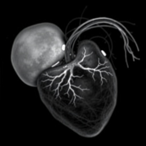
Image Credits: AI Generated
DOI: https://doi.org/10.1007/s00247-025-06306-2
Keywords: fetal congenital heart disease, magnetic resonance imaging, prenatal diagnosis, cardiac function, non-invasive imaging
