In a groundbreaking advancement that could revolutionize breast cancer screening, researchers at Mass General Brigham have demonstrated the technical feasibility of utilizing ultra-low field (ULF) magnetic resonance imaging (MRI) technology for breast imaging. This pioneering work explores the potential of ULF MRI to become a lower-cost, more accessible alternative to traditional screening modalities, which may dismantle existing barriers to early detection and improve patient experiences. Published recently in the esteemed journal Scientific Reports, the study presents compelling initial evidence that this emerging technology can visualize essential breast structures without the drawbacks commonly associated with mammography and conventional MRI.
Traditional breast cancer screening, predominantly conducted via mammography, poses several challenges. Standard mammographic procedures require breast compression, which patients often describe as uncomfortable or even painful. Moreover, mammography employs ionizing radiation, raising concerns about cumulative exposure, especially with routine screening recommendations that currently advise women aged 40 to 74 years to participate regularly. While standard high-field MRI is occasionally utilized for high-risk patients, its prohibitive cost and limited availability restrict its application in general population screening. Against this backdrop, ULF MRI emerges as a promising innovation, boasting acquisition costs that are less than 5% of conventional MRI systems and markedly reduced operational expenses.
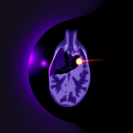
The Mass General Brigham study rigorously tested ULF MRI on a modest cohort of 14 women, spanning a diverse clinical history including 11 healthy participants, two with a previous breast cancer diagnosis, and one harboring a benign breast mass. Despite the extremely low magnetic field strength relative to traditional high-field MRIs, the scans revealed that three expert radiologists could consistently identify critical breast tissue features. They successfully distinguished fibroglandular tissue from adipose tissue, an essential capability for detecting abnormalities. The study notes that minor discrepancies among interpretations likely reflect the nascent stage of ULF MRI use in breast imaging and anticipate improvements through increased training and experience.
One of the most compelling advantages of ULF MRI is its non-reliance on ionizing radiation or intravenous contrast agents, differentiating it fundamentally from both mammography and conventional MRI methods. This aspect not only supports patient safety by reducing radiation risk but also broadens the screening applicability to populations contraindicated for contrast use. The non-invasive nature and the elimination of compression could enhance patient comfort, potentially increasing screening adherence among populations hesitant to undergo traditional procedures.
Developed by a multidisciplinary team led by Matthew Rosen, PhD, an associate professor of Radiology and director of the Low Field MRI Laboratory at the Athinoula A. Martinos Center for Biomedical Imaging, the study exemplifies the integration of engineering marvels with clinical needs. The ULF MRI devices used generate magnetic fields at a fraction of the strength of conventional MRI scanners, presenting a unique challenge in image resolution and signal-to-noise ratios. Nonetheless, the preliminary data reveal that these technical challenges can be surmounted, allowing meaningful breast tissue visualization that could support diagnostic and screening workflows.
Co-lead author Neha Koonjoo, PhD, emphasized the transformative potential of the technology, highlighting that ULF MRI can detect essential anatomical features and some abnormalities without exposing patients to radiation or contrast agents. This suggests a paradigm shift wherein breast screening may evolve into a safer and more patient-centric process while maintaining diagnostic efficacy. If scaled and validated in larger clinical cohorts, ULF MRI might serve either as a primary screening tool or as an adjunct, complementing existing modalities and stratifying patients by risk more effectively.
Kathryn E. Keenan, PhD, representing the U.S. National Institute of Standards and Technology and co-principal investigator of the project, acknowledged that achieving visible breast features at ultra-low magnetic fields was uncertain prior to the study. The successful outcomes invigorate ongoing research efforts, underscoring the robust collaboration between academic health systems and federal research institutions. Together, these partnerships are pivotal to overcoming the technical barriers inherent in ULF MRI deployment and optimizing its clinical utility.
Despite these promising strides, the researchers caution that further studies involving larger populations—incorporating diverse breast pathology including benign and malignant lesions—are necessary to fully assess the diagnostic accuracy of ULF MRI. Moreover, technological advancements must continue to refine image acquisition parameters to meet the clinical resolution standards required for reliable breast cancer screening and diagnosis. These enhancements are critical to ensure that ULF MRI images deliver sufficient detail for medical decision-making comparable to or surpassing current methods.
The team anticipates that the insights gleaned from this early feasibility study will inform essential engineering redesigns intended to improve image quality, scanning comfort, and ease of operation. Such improvements could lower logistical and economic barriers that currently restrict widespread breast cancer screening, directly benefiting underserved and resource-limited communities. By bringing breast screening closer to patients in various care settings—including primary care clinics and remote locations—ULF MRI has the potential to democratize access to early detection.
Further, by reducing reliance on contrast injections and radiation, screening workflows could become more streamlined, less costly, and more acceptable to patients wary or unable to undergo traditional imaging. The lower hardware and maintenance costs of ULF MRI systems could offer healthcare providers increased flexibility, enabling expanded outreach efforts and more frequent screenings, which are vital for early cancer detection and improved clinical outcomes.
This compelling research, supported by grants from the National Institutes of Health, the Kiyomi and Ed Baird MGH Research Scholar award, the German-American Fulbright Commission, the National Institute of Standards and Technology, and the U.S. Department of Commerce, illuminates a future where precision medical imaging is accessible to all. As the field of low-field MRI technologies advances, patients and clinicians alike may witness the advent of safer, more tolerable, and economically feasible breast cancer screening options that ultimately save lives.
The enthusiastic response from the scientific community and clinical stakeholders underscores the importance of innovative imaging solutions for public health. By building upon this proof-of-principle work, the convergence of bioengineering, radiology, and clinical practice heralds a new chapter in oncologic imaging: one characterized by patient comfort, safety, affordability, and equity.
Subject of Research: People
Article Title: Breast imaging with ultra-low field MRI
News Publication Date: 19-Feb-2026
Web References:
10.1038/s41598-026-37130-9
References: Shen S, Koonjoo N, et al. “Breast imaging with ultra-low field MRI.” Scientific Reports, DOI: 10.1038/s41598-026-37130-9
Keywords: Imaging, Mammography
