Ovarian cancer remains one of the deadliest gynecologic malignancies worldwide, primarily due to its stealthy nature and the difficulty in detecting it at early, more treatable stages. Traditional screening methods, including serum biomarkers like CA-125 and imaging techniques such as transvaginal ultrasound, often fall short of identifying cancer before it advances. Groundbreaking research over the past decade, however, has redirected scientific focus from the ovaries themselves to the fallopian tubes, which are now recognized as a primary site where many aggressive ovarian cancers originate. This paradigm shift has underscored the urgent need for innovative diagnostic tools tailored to probe the intricate anatomy of the fallopian tubes with the precision required for early disease detection.
In an exciting advancement described in a recent publication in Biophotonics Discovery, a team of bioengineers and clinicians unveiled a next-generation endoscopic device tailored explicitly for imaging the interior of the fallopian tubes while concurrently capturing cellular samples. This device, coined the Cell-Acquiring Fallopian Endoscope (CAFE), embodies a significant leap in diagnostic technology by miniaturizing imaging capabilities to navigate the submillimeter luminal space of the tubes and integrate cell collection mechanisms within a single probe. The CAFE system leverages advanced optical components and refined mechanical design to overcome the anatomical challenges that have historically impeded effective falloposcopic examination.
High-grade serous carcinomas (HGSC) constitute the most prevalent and lethal subtype of ovarian cancer. Increasingly, the scientific consensus supports the hypothesis that these malignancies emerge not from the ovarian surface epithelium but from early precursor lesions confined to the secretory epithelial cells lining the fallopian tubes. These premalignant lesions, or serous tubal intraepithelial carcinomas (STICs), can remain localized for years before disseminating to the ovary and peritoneal cavity. This temporal window presents a critical opportunity for interception and early diagnosis if targeted imaging and molecular sampling can be successfully implemented.
One of the fundamental obstacles in imaging the fallopian tubes relates to their diminutive diameter, often less than one millimeter, coupled with highly folded, flexible walls. Conventional falloposcopes offered limited field of view, mechanical rigidity that compromised navigation, and lacked the sophistication needed for simultaneous cell acquisition. The newly engineered CAFE device, crafted in close consultation with surgical and gynecologic oncology specialists, addresses these shortcomings by adopting a multifiber optical bundle with higher density and developing a bespoke close-focus lens system. This optical design permits crisp imaging at distances mere hundreds of microns from the tissue surface, delivering unprecedented resolution under both white-light and fluorescence illumination.
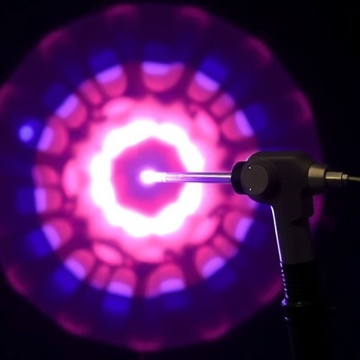
The optical imaging modalities integrated into the CAFE encompass white-light reflectance for anatomical orientation, blue-light reflectance to discern subtle textural variations, and 405 nm autofluorescence imaging which exploits endogenous fluorophores within cellular and extracellular components. Upon illumination, pathologic tissue exhibits altered metabolic and structural characteristics that modulate emitted fluorescence; these shifts are measurable and can differentiate between normal and abnormal epithelium. The high sensitivity of the fluorescence imaging optimized in CAFE enables rapid acquisition of diagnostically relevant signals even with shutter speeds as low as 100 milliseconds, minimizing motion artifacts and patient discomfort.
Beyond imaging, the innovative mechanical architecture includes a smooth, scoop-like cell collection system housed within an ultrathin working channel. Unlike conventional biopsy techniques that utilize exposed wires to scrape tissue—posing risks of injury and inflammation—the CAFE’s gentle epithelial cell capture mechanism minimizes trauma while acquiring sufficient cellular yield for downstream cytological and molecular assays. Tests in freshly excised human fallopian tubes confirmed the endoscope’s capacity to harvest tens to hundreds of thousands of epithelial cells per sampling event, an ample quantity for sophisticated diagnostic analyses such as genomic sequencing, proteomics, or immunocytochemistry aimed at early neoplastic transformation markers.
Safety and device robustness were rigorously evaluated through compliance testing against electrical and laser safety standards, as well as sterilization protocols. Tissue samples post-imaging and sampling revealed no discernible damage or structural compromise, highlighting the potential of CAFE for repeated use in clinical or surveillance settings. The system’s mechanical flexibility and compatibility with guidewire navigation facilitate seamless advancement through the convoluted fallopian tubes, a feature critical for in vivo applications and patient tolerability.
Critically, the researchers employed quantitative metrics derived from multispectral imaging data rather than relying solely on absolute fluorescence intensities. Ratios of reflectance to fluorescence signals, along with inter-channel color analyses of white-light images, were consistent between corresponding tubes within individual patients, underscoring the reliability and reproducibility of optical signatures representative of tissue health or pathology. These analytical advancements set the stage for establishing robust diagnostic criteria in larger cohorts comprising precursor lesions and early malignancies.
This research stands at the forefront of efforts to extend minimally invasive diagnostic surveillance to individuals at heightened risk for ovarian cancer, including carriers of germline BRCA1 and BRCA2 mutations. Currently, such high-risk patients frequently undergo prophylactic salpingo-oophorectomy, a surgery with significant implications for fertility and hormonal balance. The prospect of non-destructive, periodic fallopian tube assessments using devices like CAFE represents a transformative strategy for monitoring pre-cancerous changes, thereby personalizing risk management and possibly deferring invasive interventions.
By fusing highly detailed optical imaging with a non-traumatic method for cell sampling in a device under one millimeter in diameter, the Cell-Acquiring Fallopian Endoscope illuminates a path toward earlier detection and more precise characterization of where ovarian cancer originates. Continued refinement, clinical trials incorporating pathological tissues, and integration with molecular diagnostics will determine its ultimate role in gynecologic oncology. Nonetheless, this pioneering technology exemplifies how multidisciplinary innovation—melding optics, engineering, and clinical insight—can redefine paradigms in cancer detection and prevention.
In summary, the development of the CAFE system could represent a watershed moment in ovarian cancer diagnostics. Its ability to probe the fallopian tubes’ complex architecture with fine-scale resolution, paired with an elegantly simple yet effective cell collection method, promises to enhance understanding of the earliest carcinogenic processes. This innovation holds the potential to improve outcomes dramatically by shifting ovarian cancer detection to a stage where curative treatments are far more achievable, fundamentally altering the landscape of women’s health diagnostics in the decades to come.
Subject of Research: Cells
Article Title: Improved endoscope for imaging and cell collection in the fallopian tubes
News Publication Date: 17-Mar-2026
Web References:
- https://www.spiedigitallibrary.org/journals/biophotonics-discovery/volume-3/issue-02/025001/Improved-endoscope-for-imaging-and-cell-collection-in-the-fallopian/10.1117/1.BIOS.3.2.025001.full
- http://dx.doi.org/10.1117/1.BIOS.3.2.025001
References:
Gálvez, D., et al. (2026). Improved endoscope for imaging and cell collection in the fallopian tubes. Biophotonics Discovery, 3(2), 025001. https://doi.org/10.1117/1.BIOS.3.2.025001
Image Credits: D. Gálvez et al
Keywords: Ovarian cancer, Cancer cells, Fallopian tube imaging, Endoscope, Cell collection, Fluorescence imaging, Early cancer detection, High-grade serous carcinoma, Gynecologic oncology, Optical diagnostics
