In an exciting development for the field of neuroscience, researchers have made significant strides in the identification and diagnosis of cognitive impairments in patients with Parkinson’s disease using advanced imaging techniques. The study, conducted by Zeng, Liang, Guo, et al., focuses on the integration of radiomics features derived from hippocampal functional imaging. This innovative approach not only sheds light on the underlying neurological mechanisms involved in Parkinson’s disease but also opens up new pathways for earlier and more accurate diagnoses.
Parkinson’s disease, a neurodegenerative disorder that affects millions worldwide, is characterized by a range of cognitive impairments alongside its more recognizable motor symptoms. Patients often suffer from issues like memory loss, difficulty with attention, and changes in executive functioning. The severity and onset of these cognitive symptoms can vary widely among individuals, complicating diagnosis and management. Traditional diagnostic methods mainly rely on clinical evaluations, which can sometimes overlook subtle cognitive deficits until they have progressed to more advanced stages.
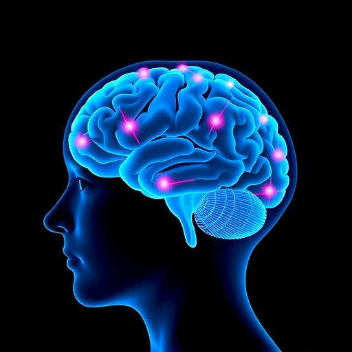
The research conducted by Zeng and colleagues seeks to address these limitations. By utilizing functional magnetic resonance imaging (fMRI) to assess hippocampal activity, the researchers were able to extract a wealth of data regarding brain functionality and connectivity. The hippocampus, a critical region associated with memory and learning, has been shown to exhibit changes in activity patterns in individuals with Parkinson’s disease. This study focuses on quantifying those changes through radiomic analyses—an emerging field that employs high-dimensional feature extraction techniques to analyze complex biomedical images.
One of the most striking findings of the study is the correlation between specific radiomic features and cognitive impairment as assessed by standard neuropsychological tests. The researchers identified unique patterns in hippocampal activity that were significantly associated with varying degrees of cognitive decline in the study group. This correlation signifies that functional imaging may serve as a potential biomarker for identifying cognitive impairment in Parkinson’s disease patients, marking a shift towards more objective diagnostic criteria grounded in neurobiological metrics.
Moreover, the study emphasizes the potential of radiomics in capturing the heterogeneity of disease expression among patients. Since Parkinson’s disease manifests differently in each individual, relying solely on clinical assessments can lead to misdiagnosis or delayed treatment. The implementation of radiomic features allows for a more nuanced understanding of how disease impacts cognitive function, paving the way for personalized medicine approaches that could be tailored to individual patients based on their specific cognitive profiles.
Another fascinating aspect of the research is the application of machine learning techniques to analyze the radiomic data. The team employed algorithms that can process immense data sets resulting from the imaging studies, identifying patterns that might not be readily apparent to human observers. This computational approach highlights the growing intersection between machine learning and neuroscience, where technology is harnessed to unveil intricate relationships within biological data. The ability to predict cognitive impairment with high accuracy based on machine learning models represents a paradigm shift that could enhance clinical decision-making significantly.
As with all pioneering research, this study faces some challenges and limitations. Among them is the necessity for further validation across larger and more diverse cohorts. While the initial findings are promising, they need to be confirmed in broader populations to ensure they are robust and generalizable. Additionally, the integration of radiomic features into routine clinical practice will require substantial efforts in training practitioners and developing protocols that can seamlessly incorporate these advanced imaging techniques.
The researchers advocate for further interdisciplinary collaboration, emphasizing the importance of merging radiology, neurology, and computational sciences to foster breakthroughs in diagnosing neurodegenerative diseases. Future research should aim to explore the applicability of radiomic features in other aspects of Parkinson’s disease, such as therapy response monitoring and progression assessment. This could ultimately lead to a comprehensive framework that utilizes imaging data to not only diagnose but also manage the disease more effectively.
Patients themselves stand to benefit from the advances depicted in this study. As diagnoses become more accurate and personalized, treatment plans can be fine-tuned to address specific cognitive deficits. Tailoring interventions based on a patient’s cognitive profile enables healthcare providers to allocate resources more efficiently and improve quality of life for those affected by Parkinson’s disease.
Additionally, the implications of this research extend beyond patients with Parkinson’s disease. The methodologies developed by Zeng and colleagues could potentially inform research into other neurodegenerative disorders that present with cognitive impairments, such as Alzheimer’s disease or frontotemporal dementia. By refining radiomic analysis techniques, researchers hope to uncover commonalities and divergences in brain mechanisms across various conditions, promoting a deeper understanding of neurodegeneration as a whole.
In conclusion, the work by Zeng, Liang, Guo, and their team represents a groundbreaking advancement in the field of cognitive neuroscience. Their findings underscore the vital role that advanced imaging techniques and radiomics can play in enhancing diagnostic accuracy for cognitive impairments associated with Parkinson’s disease. As the journey to better understand and manage neurodegenerative conditions continues, this research paves the way for a future where cognitive assessment is more precise, personalized, and ultimately, more effective in safeguarding the quality of life for patients suffering from these conditions.
The promise of these findings lies not just in the science itself but in the potential for practical application in clinical settings, where early diagnosis and tailored treatment strategies could significantly alter the trajectory of disease progression in many patients. As the field moves forward, the intersection of neuroscience, technology, and patient care takes a significant leap towards realizing a more hopeful future for those confronting the challenges of cognitive decline in Parkinson’s disease.
Subject of Research: Hippocampal functional imaging-derived radiomics features for diagnosing cognitively impaired patients with Parkinson’s disease.
Article Title: Hippocampal functional imaging-derived radiomics features for diagnosing cognitively impaired patients with Parkinson’s disease.
Article References:
Zeng, W., Liang, X., Guo, J. et al. Hippocampal functional imaging-derived radiomics features for diagnosing cognitively impaired patients with Parkinson’s disease.
BMC Neurosci 26, 27 (2025). https://doi.org/10.1186/s12868-025-00938-8
Image Credits: AI Generated
DOI: https://doi.org/10.1186/s12868-025-00938-8
Keywords: Parkinson’s disease, cognitive impairment, hippocampus, radiomics, functional imaging, machine learning, neurodegeneration, personalized medicine, diagnostic accuracy, advanced imaging techniques, interdisciplinary collaboration, neuropsychology, treatment strategies.
