In the relentless fight against pancreatic cancer, one of the deadliest malignancies with notoriously poor survival rates, a groundbreaking study has emerged to offer new hope. Scientists have developed an innovative prognostic model that merges advanced radiomics with cutting-edge 3D deep learning techniques, harnessing the power of medical imaging and artificial intelligence to predict patient outcomes more accurately. This fusion approach promises personalized treatment strategies that could significantly change the landscape of pancreatic cancer care.
Pancreatic cancer remains a formidable challenge due to its rapid progression and late diagnosis, which often leaves clinicians with limited tools for predicting how individual patients will fare. Conventional methods rely heavily on clinical judgment and basic imaging assessments, typically falling short in prognostic detail. Recognizing this gap, researchers embarked on a rigorous investigation spanning a decade, analyzing data drawn from 880 patients treated across two major hospitals between 2013 and 2023.
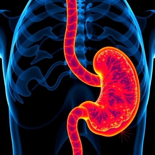
Central to this study was the use of portal venous phase contrast-enhanced computed tomography (CT) scans, which provide detailed visualizations of the pancreatic tumors. Two experienced physicians meticulously delineated tumor regions of interest (ROIs), ensuring high-quality input data integral for precise feature extraction. From these ROIs, an extensive set of 1,037 radiomic features was computed, encompassing a vast array of quantitative descriptors such as texture, shape, and intensity metrics that describe tumor heterogeneity invisible to the naked eye.
Given the overwhelming volume and complexity of these features, the research team employed principal component analysis (PCA) for dimensionality reduction, helping to distill the most critical patterns. LASSO regression further fine-tuned this selection, isolating variables most strongly associated with survival outcomes. This rigorous feature selection process ensured that the resulting radiomics model would robustly handle the prediction of overall survival while accounting for the censored nature of clinical survival data.
Parallel to the radiomics approach, the investigators developed a 3D-DenseNet deep learning model designed to extract sophisticated imaging features directly from the ROI-based 3D image volumes. DenseNet architecture, known for efficient feature reuse and gradient flow, was leveraged to capture nuanced spatial relationships within the tumor, beyond traditional handcrafted features. This neural network was trained to predict survival status at distinct time points—1-year, 2-year, and 3-year—offering temporal granularity vital for clinical decision-making.
Crucially, the innovation lies in the fusion of these two distinct modalities. The study integrated radiomic features, deep learning outputs, and baseline clinical data into composite models using several machine learning classifiers including logistic regression, random forest, support vector machine, and decision tree algorithms. The fusion was framed as a binary classification task, aiming to determine survival status at targeted temporal milestones, a practical scenario for oncologists tailoring treatment plans.
Performance evaluation revealed that while each unimodal model exhibited strong predictive capabilities, the fusion model consistently outshone them. In the test cohort, the fusion model achieved remarkable area under the curve (AUC) values—0.87 for 1-year, 0.92 for 2-year, and an impressive 0.94 for 3-year survival prediction. Accuracies also peaked at 0.84, 0.86, and 0.89 respectively, marking substantial improvements over the radiomics and 3D-DenseNet models alone.
A remarkable aspect of the study was the exploration of feature contributions within the fusion model, unveiling that deep learning features extracted via the 3D-DenseNet had the most influential role in survival predictions. Radiomic features carried significant weight as well, while clinical variables complemented these imaging-derived data, collectively enabling a nuanced assessment of disease prognosis that surpasses traditional standards.
The authors demonstrated the clinical utility of their model by stratifying patients into high-risk and low-risk categories based on the fusion model’s predictions. Kaplan-Meier survival analyses and Log-rank tests underscored statistically significant differences in overall survival between these groups, emphasizing the model’s potential to guide personalized therapeutic strategies and optimize resource allocation in clinical oncology.
This study represents a significant leap forward in oncologic imaging and machine learning integration, positioning radiomics and 3D deep learning not as competing entities but as synergistic tools for enhanced prognostication. By blending detailed tumor characterization with powerful computational pattern recognition, the fusion model embodies the next frontier of precision medicine in pancreatic cancer.
Moreover, the methodological rigor and multi-institutional nature of the dataset lend robustness and generalizability to the findings, suggesting that such fusion models could be adapted and validated across diverse clinical settings. Future efforts may aim to incorporate additional biomarkers, such as genomic or serum-based data, further enriching predictive power and mechanistic insights.
The implications for patient care are profound. Accurate survival predictions enable clinicians to tailor interventions, balancing aggressive treatments with palliative care when appropriate, thereby improving quality of life and optimizing clinical outcomes. Furthermore, such models can inform clinical trial designs by identifying suitable candidates who might benefit most from investigational therapies.
In conclusion, the fusion of radiomics and 3D deep learning holds immense promise for transforming pancreatic cancer prognosis. This study illuminates a path toward harnessing complex image-derived data with artificial intelligence to unlock predictive insights previously unattainable through conventional means. As computational methods continue to evolve, their integration into clinical oncology workflows becomes imperative for advancing personalized medicine.
The development of this fusion prognostic model heralds a paradigm shift, demonstrating that the convergence of technology and medicine can yield powerful new tools to confront one of the most lethal cancer types. With continued research and clinical validation, such innovations may soon move from the pages of scientific journals into everyday clinical practice, offering renewed hope for patients battling pancreatic cancer worldwide.
Subject of Research: Prognostic prediction models in pancreatic cancer combining radiomics and 3D deep learning approaches.
Article Title: Development of a radiomics-3D deep learning fusion model for prognostic prediction in pancreatic cancer
Article References:
Dou, Z., Lu, C., Shen, X. et al. Development of a radiomics-3D deep learning fusion model for prognostic prediction in pancreatic cancer. BMC Cancer 25, 1612 (2025). https://doi.org/10.1186/s12885-025-14889-0
Image Credits: Scienmag.com
DOI: https://doi.org/10.1186/s12885-025-14889-0
