A New AI-Driven Approach Reveals Early Signs of Progression in Fibrotic Interstitial Lung Disease
In a groundbreaking study published in the American Journal of Respiratory and Critical Care Medicine, researchers from National Jewish Health have demonstrated that subtle yet measurable increases in lung fibrosis, detected through artificial intelligence (AI)-enhanced CT imaging, are closely tied to disease progression and patient survival in fibrotic interstitial lung disease (ILD). This pioneering work offers hope for earlier detection and intervention in diseases like idiopathic pulmonary fibrosis (IPF), where timely treatment can dramatically alter patient outcomes.
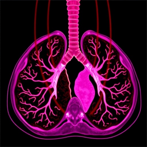
Fibrotic ILDs encompass a group of chronic disorders characterized by progressive scarring within the lung interstitium, which impairs respiratory function and ultimately leads to respiratory failure. Traditionally, clinicians rely on symptomatic assessments, lung function tests such as forced vital capacity (FVC), and the qualitative interpretation of high-resolution computed tomography (HRCT) scans by radiologists to monitor disease progression. However, this approach is often hampered by subjective biases and high variability, particularly when evaluating minute changes over successive scans.
The National Jewish Health team tackled these limitations by utilizing a sophisticated deep learning method known as data-driven textural analysis (DTA). This AI-based tool quantifies the extent of fibrosis within the lung parenchyma by analyzing detailed CT image textures, enabling objective, reproducible measurements of subtle disease evolution. Importantly, the research focused on comparing fibrosis scores derived from CT scans taken one year apart, revealing that even modest increases are prognostically significant.
Dr. Matthew Koslow, the lead pulmonologist on the study, explained the clinical relevance: “Our findings reveal that a mere 5% or greater increase in fibrosis quantified by DTA corresponds to more than double the risk of death or lung transplantation within the following year. Remarkably, this signal is most pronounced in patients whose disease was initially less severe, highlighting a critical window in which early intervention might have the greatest therapeutic impact.”
Beyond correlations with survival, the increase in DTA fibrosis scores also strongly predicted accelerated declines in lung function parameters, affirming that radiologic progression mirrors clinical deterioration. These insights provide a promising new biomarker that can supplement lung function testing to more accurately monitor fibrotic ILD progression.
The statistical rigor of the study was bolstered by the expertise of biostatistician Dr. David Baraghoshi, who underscored how combining quantitative imaging with robust modeling can illuminate patterns previously obscured by noise and subjectivity. By linking incremental changes in fibrosis extent over time to longitudinal health outcomes, the team showcased the powerful prognostic value embedded in imaging data that conventional assessment methods struggle to extract.
Validation using independent data from the Pulmonary Fibrosis Foundation Patient Registry further underscored the generalizability and robustness of the AI-driven fibrosis quantification approach, suggesting widespread applicability across diverse patient populations and clinical settings.
This innovation holds transformative potential for both clinical practice and research. In clinical trials, quantitative CT-derived fibrosis scores may serve as sensitive, objective endpoints, increasing the power to detect treatment effects over shorter durations. Clinicians could leverage serial DTA scoring to stratify patients by risk more precisely, optimizing personalized treatment strategies and potentially improving survival rates.
Although antifibrotic therapies have become the cornerstone of IPF management, challenges remain in identifying which patients will progress rapidly and thus warrant aggressive treatment versus those who remain stable. The integration of AI-based CT analysis into routine workflows could fill this critical knowledge gap, enabling timely clinical decisions grounded in reproducible quantitative data rather than subjective interpretation alone.
National Jewish Health continues its tradition of pioneering respiratory medicine research, with efforts such as this study marking a new era where AI seamlessly augments human expertise in battling complex chronic lung diseases. Through relentless innovation, the hope is to not only extend lives but also preserve quality of life for thousands affected by fibrotic ILDs worldwide.
With the confluence of machine learning, clinical insight, and advanced imaging, this research exemplifies how emerging technologies are reshaping diagnostic paradigms. As the field moves forward, it is anticipated that AI-driven quantitative imaging will become an indispensable element of precision medicine in pulmonary care.
For patients, caregivers, and clinicians alike, these findings signal a promising advance toward earlier detection, improved monitoring, and better-targeted therapies in fibrotic lung disease—offering renewed optimism in the face of a formidable clinical challenge.
Subject of Research: Quantitative assessment of fibrosis progression in fibrotic interstitial lung disease using AI-based CT imaging
Article Title: (Not specified in the provided text)
News Publication Date: October 1, 2025
Web References:
– Article: https://www.atsjournals.org/doi/abs/10.1164/rccm.202503-0535OC
– National Jewish Health: http://njhealth.org
– Media resources: https://www.nationaljewish.org/about/news/media-resources
References:
American Journal of Respiratory and Critical Care Medicine, DOI: 10.1164/rccm.202503-0535OC
Image Credits: Not provided
Keywords: Fibrotic interstitial lung disease, idiopathic pulmonary fibrosis, AI, deep learning, data-driven textural analysis, lung fibrosis, quantitative imaging, CT scan, disease progression, lung function decline, clinical outcomes, pulmonary fibrosis
