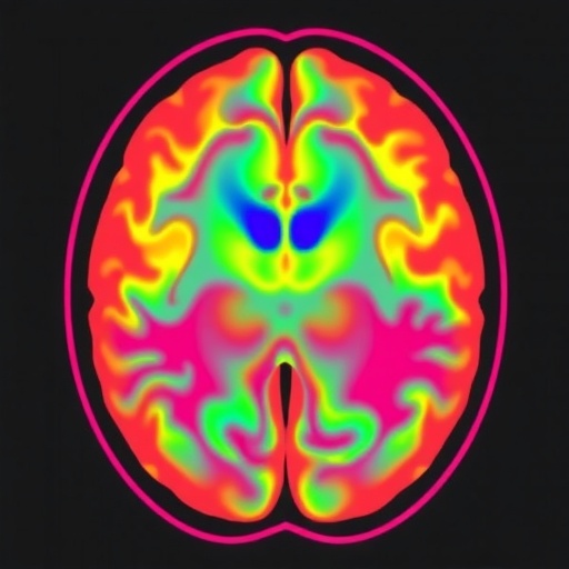
In recent years, magnetic resonance imaging (MRI) has undergone a transformative evolution, especially concerning the developing brain in pediatric populations. The latest advancements in this domain are reshaping our understanding of neurodevelopmental processes and promise profound implications for diagnosing and managing pediatric neurological disorders. The complexity inherent to the rapidly changing pediatric brain necessitates imaging technologies that can capture not only anatomical detail but also dynamic physiological and metabolic processes. This wave of innovation in MRI technology is enabling clinicians and researchers to peer deeper into the intricacies of brain maturation than ever before.
One of the most striking advancements in pediatric brain MRI lies in the enhancement of resolution and the speed at which images can be acquired. Traditional MRI scans required lengthy sessions, posing significant challenges with young patients who may struggle to remain still. Current developments in rapid imaging sequences and motion correction algorithms have dramatically reduced scan times while maintaining or improving image clarity. This progress not only enhances patient compliance but also opens doors to functional imaging and longitudinal studies that were previously impractical for pediatric subjects.
These technological leaps are integrated with sophisticated quantitative MRI techniques that yield biomarkers capable of characterizing neural tissue microstructure, myelination, and cortical development. For instance, diffusion tensor imaging (DTI), alongside newer diffusion MRI methods, is revealing white matter tract integrity with unprecedented detail. Such granular insights are indispensable for understanding neurodevelopmental disorders such as autism spectrum disorder and cerebral palsy, conditions where altered connectivity patterns play a critical role. Quantitative susceptibility mapping (QSM) and arterial spin labeling (ASL) perfusion imaging further enrich the spectrum of measurable physiological parameters.
Beyond structural and microstructural imaging, functional MRI (fMRI) technologies in pediatric settings have reached new heights. The advent of resting-state fMRI protocols tailored for children enables the mapping of intrinsic brain networks without requiring active task performance—overcoming a major hurdle in pediatric neuroimaging. These resting-state studies have elucidated the timeline and topology of brain network maturation, supporting the notion of dynamic rewiring during childhood and adolescence. This functional perspective is critical for interpreting typical and atypical cognitive development.
Another frontier involves the integration of advanced MRI with machine learning algorithms that analyze vast datasets from pediatric cohorts. These computational approaches can detect subtle patterns and predict developmental trajectories or outcomes with greater accuracy than traditional imaging analyses. By harnessing artificial intelligence, researchers are building predictive models that assist clinicians in early intervention planning and personalized therapeutic strategies, tailored to the unique neurodevelopmental profile of each child.
Challenges remain, however. The developing brain is highly heterogeneous across different age ranges, demanding age-specific imaging protocols and normative datasets. Moreover, sedation risks and ethical considerations must be meticulously managed, necessitating continuous innovation in non-invasive and child-friendly imaging techniques. Researchers are also focusing on creating open-access pediatric brain MRI repositories that encourage collaborative efforts and accelerate the pace of discovery.
One remarkable advancement is the use of multi-parametric MRI that combines structural, functional, and metabolic imaging within a single session. This multifaceted approach permits comprehensive characterization of the brain environment, providing a holistic understanding of neurodevelopmental health or pathology. Such integrative scans facilitate the differentiation of transient developmental anomalies from permanent abnormalities, improving diagnostic precision.
The clinical implications of these advances cannot be overstated. Disorders such as neonatal hypoxic-ischemic encephalopathy, early onset epilepsy, and congenital brain malformations are better characterized and monitored using these sophisticated MRI techniques. Early detection of subtle changes in the developing brain promises timely therapeutic interventions that may alter trajectories toward improved outcomes. Furthermore, MRI is playing a pivotal role in the assessment of treatment response and long-term follow-up in pediatric neuro-oncology.
Research is increasingly focused on optimizing MRI sequences to be as safe as possible, reducing radiofrequency energy deposition and acoustic noise levels, factors especially significant for infant scanning. Innovations such as silent MRI and low-field portable scanners enhance accessibility and tolerability of brain imaging, especially in neonatal intensive care settings. This democratization of advanced neuroimaging tools extends their reach beyond tertiary care centers into community hospitals.
On the horizon, hybrid imaging modalities combining MRI with other technologies, such as positron emission tomography (PET), are expected to yield synergistic benefits in understanding brain metabolism alongside anatomical changes. Although still in early stages for pediatrics, these approaches may revolutionize diagnosis and personalized medicine strategies for developmental brain disorders by providing unprecedented multi-dimensional data.
Importantly, the role of MRI extends beyond diagnosis to a powerful tool in neuroscientific research aimed at decoding the fundamental principles of brain development. High-resolution imaging is illuminating the temporal and spatial patterns of synaptic pruning, myelin maturation, and neurovascular coupling that underpin learning and cognition. Such insights fuel hypotheses for therapeutic innovation and help unravel the complex interplay between genetics, environment, and brain maturation.
Collaborations between engineers, neuroscientists, and clinicians are foundational in driving these innovations forward. Multidisciplinary teams are designing child-specific MRI coils, optimizing pulse sequences, and creating pediatric-tailored analytic pipelines. These concerted efforts ensure that technological progress translates effectively into clinical and research advancements, maximizing the impact on child health.
Moreover, ethical frameworks for pediatric MRI research are evolving to accommodate these technological strides. Emphasis on minimizing distress, optimizing informed consent, and safeguarding data privacy ensures responsible application of these powerful tools. Equitable access to advanced pediatric neuroimaging remains a priority to reduce healthcare disparities globally and ensure benefits reach all children, regardless of geography or socioeconomic status.
In summary, the ongoing advancements in magnetic resonance imaging of the developing brain represent a paradigm shift for pediatrics. The convergence of cutting-edge hardware, innovative imaging sequences, machine learning analytics, and collaborative research is illuminating the dynamic complexities of childhood brain development like never before. These innovations promise to transform both clinical practice and neuroscientific inquiry, fostering earlier diagnoses, individualized treatments, and deeper scientific understanding that holds the potential to improve countless young lives worldwide.
Subject of Research:
Advances in magnetic resonance imaging techniques focused on the developing pediatric brain and their clinical and research applications.
Article Title:
Advances in magnetic resonance imaging of the developing brain and its applications in pediatrics.
Article References:
Chen, RK., Li, MY., Zhao, ZY. et al. Advances in magnetic resonance imaging of the developing brain and its applications in pediatrics. World J Pediatr 21, 652–707 (2025). https://doi.org/10.1007/s12519-025-00905-7
Image Credits: AI Generated
DOI: July 2025


