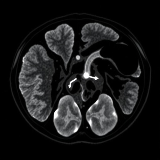
In a groundbreaking advancement at the intersection of prenatal medicine and imaging technology, researchers have unveiled a novel approach to quantifying pulmonary health in fetuses diagnosed with congenital diaphragmatic hernia (CDH). This innovative study, recently published in Pediatric Research, leverages the sophisticated magnetic resonance imaging (MRI) parameter known as T2* to non-invasively assess lung tissue oxygenation and integrity before birth. The retrospective, case-controlled pilot investigation spearheaded by Avena-Zampieri and colleagues marks a significant stride towards enhancing prenatal diagnostics and prognostication in cases burdened by one of the most formidable neonatal pulmonary malformations.
Congenital diaphragmatic hernia is a complex developmental anomaly characterized by an abnormal opening in the diaphragm, allowing abdominal organs to intrude into the thoracic cavity, consequently compromising lung formation and function. This condition often culminates in pulmonary hypoplasia and hypertension, which are primary determinants of neonatal morbidity and mortality. Current prenatal assessments rely heavily on ultrasound metrics and fetal lung volume measurements, which, while valuable, provide limited insights into the actual oxygenation status and microstructural conditions of the fetal lung parenchyma. It is within this clinical context that the study’s introduction of T2* quantification emerges as a potential game-changer.
Magnetic resonance imaging T2 relaxation time is a parameter sensitive to magnetic field inhomogeneities and tissue composition, particularly influenced by the presence of deoxygenated hemoglobin. Thus, T2 mapping serves as a surrogate marker for tissue oxygenation and microvascular characteristics. In the domain of fetal imaging, such quantification is exceptionally challenging due to fetal movement, small organ size, and the complex interplay of maternal and fetal physiology. The research team’s successful application of T2* mapping to fetal lungs represents a remarkable technical and methodological breakthrough, offering a panoramic yet detailed vista into the pulmonary environment of fetuses grappling with CDH.
This retrospective study meticulously gathered and analyzed MRI data sets from a cohort of fetuses diagnosed with CDH alongside gestational age-matched controls. Employing advanced image reconstruction and correction algorithms, the investigators extracted T2 relaxation times from defined lung regions. Their findings revealed significantly altered T2 values in the lungs of fetuses with CDH compared to controls, indicative of reduced oxygenation and altered tissue composition. Notably, these T2* deviations correlated with clinical markers of pulmonary hypoplasia, underscoring the biomarker’s potential as a prognostic tool.
The implications of this research ripple beyond mere diagnostic refinement. T2* quantification may enable clinicians to stratify disease severity with enhanced precision, tailoring in utero interventions and delivery planning accordingly. Moreover, dynamic monitoring through serial MRI scans could provide real-time insights into the progression or amelioration of pulmonary status in response to therapeutic measures, a capacity hitherto unattainable with conventional imaging modalities.
From a technical standpoint, the study surmounted numerous challenges inherent to fetal MRI. Signal acquisition was finely tuned to minimize motion artifacts, encompassing innovative gating techniques synchronized to fetal cardiac and respiratory cycles. Additionally, the quantification pipeline incorporated sophisticated modeling to differentiate tissue characteristics from confounding variables such as magnetic susceptibility variations and maternal physiology. Through these meticulous approaches, the researchers set a new benchmark for fetal imaging fidelity.
This investigation also adds a crucial layer to our fundamental understanding of CDH pathophysiology. The T2* signal shifts likely reflect microvascular remodeling and oxygen transport impairments within the compromised lungs, phenomena that are critical to the neonate’s postnatal respiratory competence. By characterizing these alterations prenatally, the study opens avenues for targeted molecular and pharmacological interventions aimed at promoting lung vascularization and maturation within the womb.
Furthermore, the pilot nature of this research underscores the necessity for larger, multi-center trials to validate and standardize T2* measurements as a routine clinical biomarker. Such efforts would need to address variability introduced by differing MRI hardware, scanning protocols, and patient populations to ensure reproducibility and broad applicability. However, the promising results reported here lay a solid foundation for these future endeavors.
In a broader context, the approach delineated by Avena-Zampieri et al. exemplifies the transformative potential of advanced quantitative MRI techniques in fetal medicine. As imaging physics and computational analytics evolve, the prospect of non-invasive, detailed tissue characterization in utero becomes increasingly attainable. This confluence of technology and clinical need heralds a new epoch in which prenatal diagnostics transcend structural assessment to embrace functional and biochemical evaluation.
The study’s integration of retrospective data further underscores how existing imaging archives can be harnessed retrospectively for novel biomarker discovery, amplifying research efficiency and scope. By mining past images with fresh analytical lenses, clinicians and scientists can unlock previously inaccessible insights without additional patient burden or resource expenditure.
Moreover, the promising correlation between pulmonary T2* values and neonatal outcomes could eventually inform parental counseling, decision-making regarding the timing and mode of delivery, as well as postnatal management strategies, including extracorporeal membrane oxygenation candidacy and ventilatory support planning. Such personalization stands to improve survival rates and long-term respiratory health in infants affected by CDH.
It is also noteworthy that this T2 quantification technique might extend beyond CDH to other fetal pulmonary conditions, including pulmonary hypoplasia secondary to oligohydramnios or skeletal dysplasias, widening the clinical impact of this imaging innovation. The versatility and specificity of T2 measurements could facilitate a comprehensive fetal lung health assessment framework.
However, several limitations warrant discussion. The relatively small sample size inherent to pilot studies restricts statistical power and generalizability. Additionally, the retrospective design imposes constraints on control over imaging timing and standardization. Prospective longitudinal studies are essential to ascertain causality and temporal dynamics of T2* changes in fetal lung development.
In conclusion, the pioneering work by Avena-Zampieri and colleagues illuminates a novel horizon in fetal medicine through pulmonary T2* quantification by MRI in congenital diaphragmatic hernia cases. By furnishing a window into the elusive microenvironment of the developing lung, this technique promises to augment diagnostic, prognostic, and therapeutic capabilities significantly. As the field advances, such sophisticated imaging biomarkers hold the potential to reshape prenatal care paradigms, ultimately improving outcomes for vulnerable neonatal populations affected by complex pulmonary pathologies.
Subject of Research: Pulmonary T2* quantification in fetuses with congenital diaphragmatic hernia
Article Title: Pulmonary T2* quantification of fetuses with congenital diaphragmatic hernia: a retrospective, case-controlled, MRI pilot study
Article References:
Avena-Zampieri, C.L., Uus, A., Egloff, A. et al. Pulmonary T2 quantification of fetuses with congenital diaphragmatic hernia: a retrospective, case-controlled, MRI pilot study. Pediatr Res* (2025). https://doi.org/10.1038/s41390-025-04091-0
DOI: https://doi.org/10.1038/s41390-025-04091-0
Image Credits: AI Generated


