In the rapidly evolving landscape of emergency medicine and battlefield triage, ultrasound imaging remains a cornerstone diagnostic tool, celebrated for its portability and minimal power requirements. Despite its critical role, the acquisition and interpretation of ultrasound images demand highly specialized skill sets, often limiting its effectiveness outside of expert hands. Addressing this challenge head-on, a groundbreaking study introduces an innovative solution: a modular, full-torso tissue-mimicking phantom designed explicitly to simulate the extended-focused assessment with sonography for trauma, or eFAST, exam. This development represents a significant leap forward in the integration of artificial intelligence (AI) with ultrasound technology, promising to revolutionize how clinicians acquire and interpret ultrasound data in high-stakes environments.
At the heart of this advancement lies the creation of a versatile tissue phantom, meticulously engineered to replicate human thoracic anatomy and physiological motion. Unlike traditional static models, this phantom dynamically simulates full thoracic motion, replicating the nuanced biomechanical behaviors encountered in real patients. This dynamic feature is crucial for training AI algorithms under realistic imaging conditions, thereby enhancing the accuracy and reliability of diagnostic outputs. The modular design allows for the insertion of simulated injuries at each critical eFAST scan site, enabling targeted training for injury detection and classification.
The significance of this innovation extends beyond mere simulation. By generating ultrasound images from the phantom, the research team successfully trained AI models to recognize and delineate specific anatomical features and pathological states. The performance metrics reported are compelling, with intersection-over-union (IOU) indices surpassing 0.80 in key tasks, and a diagnostic accuracy reaching 71.5% on blind inference datasets. These results underscore the phantom’s utility not only as a device for AI training but also as a potential standard for benchmarking AI performance in ultrasound image analysis.
This tissue-mimicking phantom addresses one of the critical bottlenecks in deploying AI-assisted ultrasound diagnostics: the scarcity of high-quality, annotated datasets. Real-world ultrasound imaging of trauma patients is often inconsistent and unpredictable, complicating the collection of standardized training data. The phantom offers a stable, reproducible source of labeled ultrasound images, facilitating the development and validation of AI models under controlled yet realistic conditions.
Moreover, the ability to simulate modular injuries enhances the phantom’s applicability for a wide range of trauma scenarios, from pneumothorax and hemothorax to abdominal hemorrhage. This versatility is particularly valuable in military medicine, where rapid and accurate triage can be lifesaving. The AI models trained on these phantom-generated datasets could assist frontline medics by providing real-time diagnostic support, potentially reducing the cognitive load and error rates associated with manual image interpretation.
Beyond AI development, the phantom holds promise as a training aid for personnel learning ultrasound examination techniques. Its lifelike anatomical features and motion emulate the challenges encountered during actual patient scans, enabling trainees to refine their skills in a risk-free environment. This aspect is crucial for broadening the competency base of emergency responders and ensuring high-quality ultrasound assessments across diverse clinical settings.
Another exciting frontier opened by this technology is the automation of ultrasound image acquisition. Current ultrasound operations involve substantial operator dependency, where image quality can vary widely based on the technician’s expertise. Incorporating AI-guided acquisition protocols, trained using data from the tissue phantom, could standardize image quality and streamline workflows. Such automation would democratize access to high-fidelity ultrasound imaging, especially in resource-limited or austere environments.
The development process of the phantom involved sophisticated materials engineering to replicate the acoustic properties of human tissues accurately. Achieving this level of biomimicry ensures that the ultrasound waves interact with the phantom in a manner comparable to real human anatomy, generating authentic imaging artefacts and reflections. This fidelity is critical for training AI models that must operate effectively in clinical environments, where signal variations and noise are the norm.
Integration of this phantom into AI research frameworks exemplifies a collaborative convergence of biomedical engineering, computer science, and clinical expertise. The approach reflects a broader trend toward creating hybrid systems that leverage physical models alongside computational algorithms to enhance medical diagnostics. As AI increasingly permeates healthcare, such tangible training tools become indispensable for bridging the gap between theoretical model performance and real-world clinical utility.
Looking forward, the implications of this technology are profound. By enabling robust AI model development and clinician training, the modular eFAST tissue phantom could accelerate the adoption of AI-augmented ultrasound diagnostics globally. This shift has the potential to improve patient outcomes dramatically, particularly in emergency and trauma care, where rapid, accurate decisions are paramount. The phantom’s modularity also invites future enhancements, including the incorporation of additional anatomical regions or pathologies, expanding its utility across various medical disciplines.
In summary, this research heralds a new era in ultrasound imaging and AI integration. Through the development of a sophisticated, anatomically accurate, and dynamically responsive tissue-mimicking phantom, the study provides a crucial platform for advancing AI-based diagnostic tools. The demonstrated success in training AI models to detect anatomical features and injury states with high precision marks a pivotal milestone. As this technology matures, it promises to empower clinicians with enhanced diagnostic capabilities, streamline ultrasound training, and ultimately improve trauma care delivery worldwide.
The potential impact on both military and civilian medical practices cannot be overstated. By bridging the gap between advanced AI algorithms and practical ultrasound imaging challenges, this modular tissue phantom represents a vital step toward smarter, faster, and more reliable triage solutions. This innovation stands as a testament to the power of interdisciplinary research and its capacity to create tools that improve medical care at its most critical junctures.
Subject of Research:
Article Title: Modular eFAST tissue phantom for AI-based ultrasound triage
Article References: Mejia, I., Hernandez Torres, S.I., Bedolla, C. et al. Modular eFAST tissue phantom for AI-based ultrasound triage. BioMed Eng OnLine 24, 115 (2025). https://doi.org/10.1186/s12938-025-01448-8
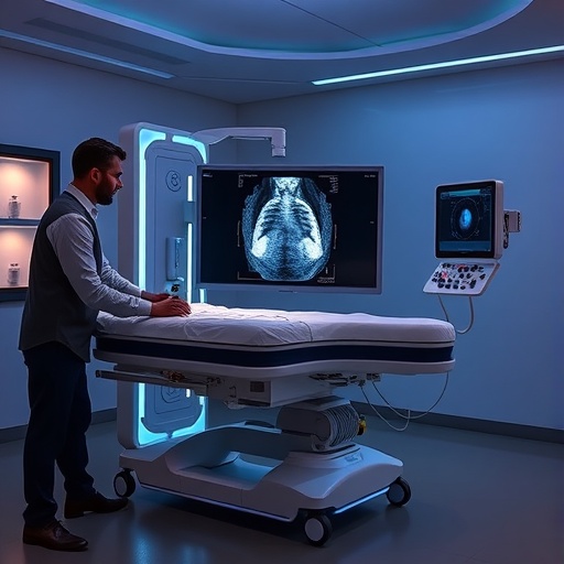
Image Credits: AI Generated
DOI: https://doi.org/10.1186/s12938-025-01448-8
