In a groundbreaking study published recently in Translational Psychiatry, researchers have unveiled significant neurobiological underpinnings linked to empathy deficits observed in patients diagnosed with Borderline Personality Disorder (BPD). Utilizing advanced functional magnetic resonance imaging (fMRI) techniques, the investigation detailed a marked reduction in activation within core brain regions responsible for empathy when individuals with BPD observed social interactions. This revelation is poised to reshape the understanding of the neural basis of social cognition impairments in personality disorders and pave the way for targeted therapeutic interventions.
Empathy, the complex ability to resonate with and understand the emotions of others, is a critical component of human social interaction. In individuals with BPD, empathy-related difficulties contribute to pervasive interpersonal dysfunction and emotional instability, often manifesting as tumultuous relationships and heightened emotional reactivity. Prior research has implicated various brain networks in empathetic processing, yet the specific neural alterations in BPD during real-time social observation remained elusive until this pioneering study.
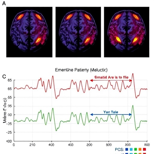
The investigative team, led by V. Flasbeck and colleagues, deployed functional MRI scans to monitor brain activity while participants engaged in tasks involving the observation of dynamic social interactions. This methodological approach allowed for the dissection of neural circuits engaged during empathy elicitation, contrasting the BPD cohort against neurotypical controls. The results compellingly indicated a hypoactivation in regions such as the anterior insula and the anterior cingulate cortex—areas critically linked to emotional awareness and affective empathy.
These findings contribute robust evidence to a neurofunctional model wherein empathy deficits in BPD correspond to subdued responsiveness within the salience network. The anterior insula, for example, operates as an integrative hub, synthesizing interoceptive signals and mediating emotional experience. Reduced activation here could underlie the blunted empathic resonance observed clinically. Concurrently, diminished anterior cingulate engagement might reflect impaired conflict monitoring and emotional regulation during social appraisal, further complicating interpersonal dynamics in affected individuals.
Beyond mapping the neural landscape, the study elegantly demonstrates the utility of fMRI paradigms in elucidating the subtleties of social cognition disruptions. The specificity of striated hypoactivation patterns in empathy-relevant regions supports the hypothesis that BPD’s clinical phenotype is partially rooted in altered brain functional connectivity and activity during social-emotional processing. This neural signature offers a promising biomarker for diagnostic refinement and patient stratification in clinical psychiatry.
Moreover, the investigation probes the implications of these neural alterations for real-world social behavior. Since empathy is pivotal for effective communication and conflict resolution, the attenuated brain responses observed may help explain the profound social difficulties characteristic of BPD. The researchers hypothesize that therapeutic strategies aiming to enhance activation in these empathy hubs—possibly through neurofeedback, cognitive-behavioral interventions, or pharmacological modulation—could ameliorate social functioning.
Methodologically, the study exemplifies rigorous fMRI data acquisition and analysis protocols, integrating high-resolution imaging with carefully validated social stimuli to evoke naturalistic empathetic responses. The use of well-matched control groups and the meticulous delineation of task conditions strengthen the reproducibility and generalizability of the results. Such methodological precision sets a precedent for future neuropsychiatric research exploring the interface of brain function and complex behavior.
Interestingly, the reduced activation patterns were consistent across varying types of social interactions presented during the tasks, indicating a pervasive deficit rather than context-dependent variability. This widespread reduction suggests fundamental disruptions in empathy circuitry rather than isolated impairments, reinforcing the conceptualization of BPD as a disorder intricately linked with emotional-processing anomalies at the neural level.
The study also discusses potential developmental trajectories that might culminate in these neural irregularities. Early adverse experiences, frequently reported in the histories of BPD patients, could result in maladaptive neuroplastic changes within empathy networks. Such insights underscore the importance of early detection and intervention, emphasizing how adverse environmental factors sculpt neurobiological vulnerability to social cognitive deficits.
Furthermore, this research invites a reevaluation of therapeutic paradigms. Conventional treatments focusing predominantly on symptomatic management might benefit from integrating neurobiological targets identified via imaging studies, fostering a more personalized medicine approach. Interventions that directly engage empathy networks, such as social cognition training, could leverage neural plasticity to restore functional capacities.
In the broader neuroscientific context, the findings align with a growing body of literature associating psychiatric disorders with specific neural circuit dysfunctions. Reducing the abstraction of mental illness to discrete behavior patterns and grounding them in neuroanatomical substrates enhances diagnostic accuracy and therapeutic efficacy. Such advances potentially reduce stigma by framing these disorders as brain-based conditions requiring comprehensive medical and psychological care.
Critically, the study recognizes its limitations, including the cross-sectional design and the moderate sample size, which may affect the extrapolation of results. Future longitudinal studies employing larger, diverse cohorts are recommended to delineate causal pathways and temporal dynamics of empathy network alterations in BPD and other related conditions.
The implications of these results extend beyond borderline personality disorder, offering a framework to interrogate empathy deficits in various neuropsychiatric illnesses, from autism spectrum disorders to schizophrenia. The nuanced understanding of empathy’s neurobiology could revolutionize social neuroscience and mental health treatment, underscoring empathy as both a clinical and scientific focal point.
In conclusion, by revealing specific reductions in empathy-related brain activations during the observation of social interactions in BPD patients, this study delivers a critical leap in the comprehension of the neurofunctional substrates underlying social dysfunctions. The integration of neuroimaging methodologies into psychiatric research heralds a new era where objective biomarkers inform both pathophysiology elucidation and personalized therapeutic design.
As the science community continues to unravel the intricate neural tapestries that orchestrate human social cognition, studies like this position empathy not merely as an abstract virtue but as a tangible neurobiological process susceptible to investigation, intervention, and ultimately healing. This work by Flasbeck et al. stands as a testament to the power of interdisciplinary collaboration in advancing mental health science.
Subject of Research: Reduced activation in empathy core regions during observation of social interactions in patients with borderline personality disorder
Article Title: Reduced activation in empathy core regions during observation of social interactions in patients with borderline personality disorder: an fMRI-study
Article References: Flasbeck, V., Enzi, B., Juckel, G. et al. Reduced activation in empathy core regions during observation of social interactions in patients with borderline personality disorder: an fMRI-study. Transl Psychiatry (2026). https://doi.org/10.1038/s41398-026-03989-5
Image Credits: AI Generated
