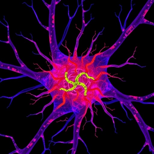
In the intricate landscape of neuronal communication, the seamless transmission of signals relies on the precise and rapid replenishment of synaptic vesicles at release sites. Groundbreaking research from Ogunmowo, Hoffmann, Patel, and colleagues, recently published in Nature Neuroscience, delves deeply into the molecular choreography that primes synaptic vesicles for replenishment, spotlighting the crucial roles of the proteins intersectin and endophilin. This study unveils how these molecules undergo liquid-liquid phase separation to form condensates, effectively orchestrating the readiness of synaptic vesicles and ensuring efficient neurotransmitter release that underpins cognition, sensation, and motor function.
Synapses, the specialized junctions where neurons communicate, depend heavily on synaptic vesicles—the membranous carriers loaded with neurotransmitters that fuse with the presynaptic membrane to release their chemical cargo. After vesicle fusion and neurotransmitter release, the rapid replenishment of release sites is vital for sustaining synaptic transmission, especially during high-frequency neuronal firing. However, the molecular underpinnings that regulate release site preparation have long remained elusive given the rapid and complex dynamics involved.
At the heart of this new research lies the discovery of the ability of intersectin and endophilin to form dynamic biomolecular condensates through liquid-liquid phase separation within presynaptic terminals. This biophysical process allows these proteins to segregate from the bulk cytoplasm without membrane encapsulation, creating concentrated hubs with distinct biochemical properties. The formation of these condensates acts as a molecular scaffold, concentrating synaptic vesicles and release machinery components in proximity to the active zone, thereby priming them for immediate release.
The study utilized advanced imaging techniques, including super-resolution microscopy and live-cell fluorescence assays, to visualize the formation and behavior of intersectin-endophilin condensates in cultured neuronal preparations. The researchers observed that these condensates rapidly assemble at the presynaptic membrane in response to neuronal activity, suggesting a tightly regulated process synchronized with synaptic demand. Notably, the condensate formation was reversible and sensitive to changes in calcium influx, emphasizing an activity-dependent mechanism that ensures synaptic vesicles are poised for replenishment only when needed.
In dissecting the molecular interactions involved, intersectin emerges as a multi-domain scaffold protein capable of binding both endophilin and synaptic vesicles. Endophilin, known for its role in membrane curvature and endocytosis, complements intersectin by contributing to the condensate’s dynamic properties and membrane interactions. Together, their cooperation fosters a microenvironment where vesicles are held in a poised state, ready to be recruited to the active zone as release sites become available.
This discovery fundamentally expands the understanding of synaptic vesicle cycling by introducing the concept that phase-separated condensates serve as a preparatory reservoir for synaptic vesicles, thereby ensuring rapid and high-fidelity neurotransmission. Previously, models focused primarily on vesicle docking and priming mediated by SNARE proteins and associated factors, but this new work places intersectin and endophilin condensates as a critical upstream regulator of release site replenishment.
Crucially, the study also explored the functional implications of disrupting these condensates. Utilizing genetic knockdown and mutant constructs that impair phase separation propensity, the researchers demonstrated a significant reduction in synaptic vesicle replenishment rates and diminished synaptic transmission efficacy. These perturbations underscored the indispensability of condensate formation in maintaining synaptic strength during repetitive stimulation, with potential implications for understanding synaptic fatigue and neurological disorders involving synaptic dysfunction.
Further analysis detailed the biophysical properties of the condensates, highlighting their sensitivity to ionic strength, pH, and post-translational modifications such as phosphorylation. These characteristics point to a sophisticated regulatory network that dynamically tunes the assembly and disassembly of intersectin-endophilin condensates in response to intracellular signaling cascades, thereby finely modulating synaptic responsiveness and plasticity.
In addition to neurons in culture, the research extended to acute brain slices and in vivo models, demonstrating that these condensates are not merely artifacts of experimental manipulation but bona fide physiological structures with conserved functions across neural circuits. This cross-validation establishes the relevance of intersectin-endophilin condensates in intact brain function and hints at their contributions to processes ranging from sensory perception to learning and memory.
The implications of these findings stretch beyond basic neuroscience, potentially informing therapeutic strategies for conditions marked by synaptic dysfunction such as epilepsy, autism spectrum disorders, and neurodegenerative diseases. Targeting the formation or stabilization of such condensates could emerge as a novel approach to bolster synaptic resilience or recalibrate aberrant neurotransmission.
Moreover, the discovery adds to the growing appreciation of biomolecular condensates as essential organizational units within cells, linking the principles of soft matter physics to biological function. Similar phase separation phenomena have been documented in diverse cellular contexts, including RNA granules, transcriptional hubs, and signaling clusters, positioning intersectin-endophilin condensates within an exciting paradigm where dynamic compartments without membranes govern key physiological processes.
The temporal precision exhibited by these condensates suggests that neurons leverage phase separation not only for spatial organization but also for rapid temporal control, enabling swift adaptation to fluctuating synaptic demands. This nuanced control likely contributes to the remarkable plasticity of the nervous system and its ability to encode information with speed and precision.
Looking ahead, the research opens numerous avenues for investigation. Deciphering how other synaptic proteins interact with intersectin and endophilin condensates, and how pathological mutations might disrupt their formation, could reveal deeper mechanistic insights. Furthermore, exploring the interplay between condensate dynamics and synaptic vesicle recycling pathways promises to illuminate the full cycle of synaptic vesicle turnover.
In sum, Ogunmowo et al. provide compelling evidence that liquid-liquid phase separation of intersectin and endophilin forms a condensate-based priming mechanism essential for replenishing synaptic vesicle release sites. This paradigm-shifting discovery enriches the understanding of synaptic transmission and offers a conceptual framework integrating biophysical principles with neuronal function—a nexus likely to inform future neuroscience research and clinical innovation alike.
Subject of Research: The molecular mechanisms by which intersectin and endophilin form biomolecular condensates to prime synaptic vesicles for rapid replenishment at neuronal release sites.
Article Title: Intersectin and endophilin condensates prime synaptic vesicles for release site replenishment.
Article References:
Ogunmowo, T.H., Hoffmann, C., Patel, C. et al. Intersectin and endophilin condensates prime synaptic vesicles for release site replenishment. Nat Neurosci (2025). https://doi.org/10.1038/s41593-025-02002-4
Image Credits: AI Generated


