A groundbreaking multicenter study spearheaded by the London Health Sciences Centre Research Institute (LHSCRI), in collaboration with the Lawson Research Institute at St. Joseph’s Health Care London and the University Health Network (UHN), has unveiled a transformative imaging methodology that significantly enhances the detection of recurrent prostate cancer. This novel approach, based on prostate-specific membrane antigen (PSMA) positron emission tomography (PET) scanning, outperforms conventional imaging techniques and correlates with improved patient survival, marking a pivotal advancement in prostate cancer diagnostics and management. The results of this extensive seven-year investigation are detailed in the latest issue of The Journal of Nuclear Medicine.
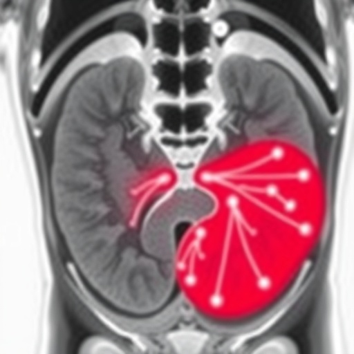
Prostate cancer recurrence poses a formidable challenge in oncological care, often eluding detection by standard imaging modalities such as bone scans and computed tomography (CT). The innovative PSMA PET scan involves the intravenous administration of a radiolabeled molecule engineered to selectively bind PSMA, a cell surface protein abundantly expressed on prostate cancer cells. This molecular targeting ensures high-contrast images by highlighting metastatic deposits with exceptional specificity and sensitivity. The study conclusively demonstrates that PSMA PET scanning identifies sites of cancer recurrence with a detection rate of approximately 70 percent, substantially surpassing historical detection rates ranging between 10 and 20 percent achieved by traditional imaging.
Dr. Glenn Bauman, a leading Radiation Oncologist at London Health Sciences Centre and Scientist at LHSCRI, emphasizes the clinical implications of this advancement: “The superior precision of PSMA PET scans allows us to detect recurrent cancer at an earlier stage and to pinpoint its exact anatomical location. This empowers clinicians to tailor therapeutic interventions specifically to the affected sites rather than resorting to systemic therapies that can be less targeted and more toxic.” This refined diagnostic capability not only enhances treatment accuracy but also enables a paradigm shift towards personalized oncology.
An essential finding from the multicenter study is the dramatic impact of PSMA PET findings on therapeutic decision-making. Approximately 50 percent of patients experienced modifications in their clinical management following PSMA PET imaging. More strikingly, nearly 90 percent of men with lesions detected via PSMA PET underwent changes in their treatment regimens, underscoring the scan’s influence on clinical practice. These treatment adaptations ranged from localized radiotherapy targeting discrete metastatic foci to the strategic initiation or alteration of systemic therapies based on precise disease burden assessments.
Beyond detection, the study highlights an observed survival advantage among patients whose management was guided by PSMA PET imaging compared to those evaluated using conventional methods. This suggests that the earlier and more accurate identification of recurrence facilitated by PSMA PET directly contributes to improved long-term outcomes, likely through enabling timely and appropriately targeted interventions. According to Dr. Ur Metser, Division Head of Molecular Imaging at UHN and Clinician Scientist at Princess Margaret Cancer Centre, “Our findings represent a monumental shift towards precision medicine in the management of recurrent prostate cancer, translating into tangible survival benefits.”
The scientific rigor of this research is further reflected in its extensive scope, enrolling thousands of men from six different hospitals across Ontario. Initiated in 2016 with the first use of PSMA PET imaging in Canada by Dr. Bauman and colleagues, the study has garnered robust funding support through Ontario Health – Cancer Care Ontario. This has facilitated comprehensive data collection, validation, and multi-institutional collaboration essential for establishing PSMA PET as a new standard of care.
Technically, PSMA PET imaging leverages positron emission tomography’s capability to detect gamma rays emitted indirectly by the radiotracer administered to patients. The tracer binds to PSMA-expressing prostate cancer cells with high affinity, accumulating in both primary and metastatic tumor sites. This accumulation generates high-resolution three-dimensional images, allowing physicians to visualize cancer spread with unparalleled clarity. Such precision imaging reduces uncertainties inherent in conventional scans and significantly improves staging accuracy.
Clinically, the advent of PSMA PET imaging addresses a critical unmet need: the detection of biochemically recurrent prostate cancer when routine scans fail to localize disease despite rising prostate-specific antigen (PSA) levels. By identifying occult metastases early, PSMA PET permits focused salvage therapies, potentially delaying or obviating the need for systemic treatments that carry higher morbidity. This diagnostic innovation thus enhances patient quality of life alongside clinical outcomes.
Moreover, the implementation of PSMA PET scanning exemplifies the interplay between molecular biology and medical imaging technologies, showcasing how targeted radiotracers can revolutionize oncological imaging. The translation of discoveries from preclinical molecular studies into clinical applications epitomizes modern precision oncology. As PSMA-targeted agents continue to be refined, future developments may include theranostic approaches that combine diagnostic imaging with targeted radionuclide therapy.
The broad adoption of PSMA PET scans as a funded healthcare service in Ontario marks a noteworthy policy achievement. It demonstrates confidence in this technology’s clinical utility and cost-effectiveness to justify public health investment. This could serve as a model for other regions seeking to integrate advanced molecular imaging into prostate cancer care pathways, promoting equitable access to cutting-edge diagnostics.
In summary, the transformative impact of PSMA PET scanning in the early detection and precise localization of prostate cancer recurrence represents a major leap forward in cancer imaging. This diagnostic tool enables oncologists to make informed, targeted treatment decisions that improve survival rates and personalize patient care. As research and clinical experience accumulate, PSMA PET promises to redefine standards of care for men battling recurrent prostate cancer, offering renewed hope and improved prognoses.
Subject of Research: People
Article Title: Not specified
News Publication Date: Not specified
Web References: The Journal of Nuclear Medicine article
Image Credits: LHSC
Keywords: Clinical medicine, Health and medicine
