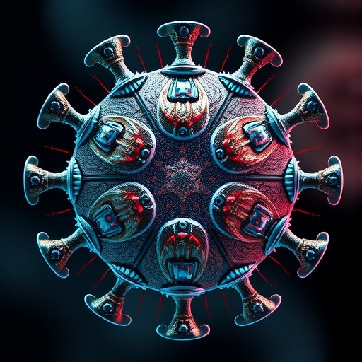
In a groundbreaking study emerging from the University of California, Riverside, physicists have unveiled an unprecedented simulation revealing how viruses meticulously assemble their protective protein shells, known as capsids, around their genetic material. These structures, often exhibiting flawless icosahedral symmetry, are fundamental to viral integrity and infectivity. The research, led by Professor Roya Zandi, transcends previous limitations by modeling the spontaneous self-assembly of larger, biologically relevant capsids, specifically T=3 and T=4 structures, which have long eluded detailed computational exploration due to their complexity.
At the heart of viral function lies the genome, the complete sequence of nucleic acids—either DNA or RNA—that codes for all viral proteins and dictates replication. Many spherical viruses encapsulate their genome within an icosahedral capsid, a geometric form consisting of twenty equilateral triangular faces arranged symmetrically. This configuration is not merely aesthetic; it optimizes the ratio between structural stability and resource economy by using multiple identical protein subunits to create a robust enclosure, efficiently protecting the viral genome from environmental damage and immune responses.
Prior research has focused primarily on smaller or artificially constrained capsid assemblies, often neglecting the dynamic interplay between genome flexibility, protein elasticity, and diffusion processes. Addressing this gap, Zandi’s team employed an innovative computational framework that simulates conformational switching in capsid subunits and models the flexible nature of viral RNA genomes. This approach provides unparalleled insight into the energetics and kinetics driving the assembly process, capturing transient intermediate states that have traditionally evaded experimental detection.
The research illuminates the paradox of viral capsid assembly: despite an inherently disordered and stochastic onset, the process culminates in highly precise, symmetrical structures. Viral genomes, often thousands of nucleotides long, serve as scaffolds that selectively recruit hundreds of protein subunits. Initially, these proteins bind in irregular and sometimes erroneous configurations; however, the intrinsic elasticity of the proteins allows mechanical stresses to fragment improper bonds, facilitating an iterative self-correction mechanism. This dynamic remodeling ultimately yields the canonical icosahedral capsid, akin to a meticulously crafted soccer ball, underscoring the self-organizing principles nature employs.
Icosahedral symmetry offers viruses a dual advantage. Structurally, it enables the assembly of a strong, closed shell with maximal internal volume relative to surface area, harboring the genome efficiently. Economically, it relies on repeating a small set of identical protein subunits, reducing the genetic coding burden on the virus. This symmetry represents an evolutionarily optimized solution for viral packaging, balancing thermodynamic stability with functional adaptability, making it a pervasive motif across numerous viral families.
Remarkably, the assembly principles discovered are widely conserved among diverse spherical viruses. While some larger viruses may require auxiliary scaffolding proteins to facilitate capsid formation, the fundamental strategy centers on elastic protein interactions that self-correct to achieve symmetry. This universality suggests that despite genetic differences, physical forces govern viral morphogenesis, offering a unifying perspective that transcends phylogenetic boundaries.
The viral genome profoundly influences capsid morphology. Its length and three-dimensional conformation, characterized by parameters such as radius of gyration, directly affect the preferred size and symmetry of the assembled capsid. Acting as a dynamic nucleation platform, the genome increases local protein concentration and stabilizes intermediate complexes, promoting efficient shell closure. The study’s focus on long RNA strands illuminates how the disordered RNA-protein condensate gradually crystallizes into an ordered shell through thermodynamically driven conformational transitions.
Experimentally, observing the fleeting intermediate stages of viral capsid assembly remains a formidable challenge. Conventional imaging techniques like cryo-electron microscopy and X-ray scattering predominantly capture static end states, obscuring dynamic growth pathways. Computational simulations, as employed in this study, bridge this gap by generating atomistic-level trajectories that expose transient assemblies and kinetic bottlenecks. This breakthrough allows for real-time visualization of the progressive formation of large capsids, including clinically significant models like hepatitis B virus, providing unprecedented mechanistic clarity.
Understanding these mechanistic details offers promising avenues for antiviral drug development. Therapeutic intervention targeting vulnerable assembly intermediates could effectively disrupt capsid formation. Potential strategies include compounds that hinder protein elasticity needed for error correction, molecules that prevent proper protein-genome interactions, or agents that stabilize improper intermediates, thus aborting formation of viable viral particles. This mechanistic targeting holds the potential to block viral replication at a crucial stage, offering specificity with potentially reduced host toxicity.
Beyond virology, the implications of this research extend into nanotechnology and medicine. The same principles guiding viral capsid assembly can be harnessed to engineer synthetic nanocontainers for drug delivery, gene therapy, and biomolecular storage. By modulating subunit elasticity and cargo properties, scientists could design customizable protein shells with precise control over size, symmetry, and cargo release dynamics. Such advancements envision custom-built nanomachines capable of targeted therapeutic delivery or responsive environmental sensing.
This interdisciplinary effort involved expertise from UC Riverside as well as collaborators from Paris-Saclay University, reflecting the global commitment to decoding viral assembly. Former graduate student Siyu Li, now faculty at Cal Poly Pomona, contributed significantly to the computational models, underscoring the role of emerging researchers in advancing foundational virology. The work was underpinned by grants from the National Science Foundation and the University of California Multicampus Research Programs and Initiatives, highlighting the importance of sustained investment in basic science.
The findings redefine how we conceptualize viral morphogenesis and open new frontiers for manipulating virus-like structures. As viruses continue to challenge global health, these insights equip researchers with advanced tools to intercept viral lifecycles and inspire biomimetic technologies. The convergence of physics, biology, and computational science charts a promising path to understanding and harnessing one of nature’s most intricate self-assembling systems.
Subject of Research: Computational simulation/modeling of viral capsid assembly
Article Title: From Disorder to Icosahedral Symmetry: How Conformation-Switching Subunits Enable RNA Virus Assembly
News Publication Date: 24-Sep-2025
Web References: DOI: 10.1126/sciadv.ady7241
Keywords: viral capsid assembly, icosahedral symmetry, RNA virus, protein self-assembly, computational simulation, conformational switching, T=3 capsid, T=4 capsid, genome-protein interaction, antiviral targets, nanotechnology, gene therapy
