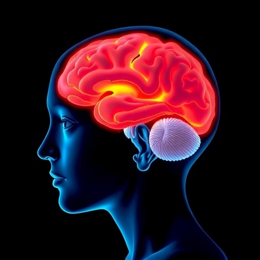
A groundbreaking new study sheds light on the intricate neural mechanisms underlying non-suicidal self-injury (NSSI) addiction in adolescents, revealing critical impairments in hippocampal function and connectivity. Published in the renowned journal BMC Psychiatry, the research employs cutting-edge resting-state functional magnetic resonance imaging (rs-fMRI) techniques to unravel how neural activity and inter-regional brain communication are altered in young individuals grappling with repetitive self-injury behaviors. These findings mark a significant leap forward in understanding the neurobiological substrates of NSSI addiction, a condition that has long mystified clinicians and neuroscientists alike.
Adolescence is a tumultuous period characterized by profound neurodevelopmental changes, and for some, this stage is marred by non-suicidal self-injury. Despite its alarming prevalence, the neural mechanisms that fuel the persistence and addictive qualities of NSSI have remained elusive. Researchers, led by Lin et al., adopted a highly sophisticated imaging approach to probe the spontaneous brain activity and functional connections in adolescents exhibiting NSSI. Their inquiry involved a carefully matched cohort of 62 participants—33 adolescents with NSSI behaviors and 29 healthy controls—ensuring that differences observed could be robustly attributed to self-injury addictive pathology rather than confounding factors.
The study’s core analytic method, amplitude of low-frequency fluctuation (ALFF), serves as a sensitive biomarker of regional spontaneous neural activity by measuring signal oscillations in specific brain areas at rest. Notably, adolescents with NSSI demonstrated marked reductions in ALFF values within both the left and right hippocampus—key regions implicated in memory processing, emotional regulation, and adaptive stress responses. Conversely, heightened activity was detected in the right supplementary motor area, suggesting altered motor planning or habit formation processes may be involved in the compulsive elements of self-injury.
Crucially, the hippocampus did not act in isolation. Functional connectivity analyses, extending from ALFF-defined regions of interest (ROIs), unveiled a pattern of disrupted network interactions. Reduced communication between the left hippocampus and several cerebral regions, including the left precuneus and temporal gyri on the right hemisphere, indicates dysfunctional neural circuits vital for integrating sensory, emotional, and cognitive data. Intriguingly, an exception was found in the form of increased connectivity between the left hippocampus and the left thalamus, which could reflect maladaptive compensatory neural network reorganization or altered relay processing critical to self-injurious behavior maintenance.
These neural aberrations are far from mere epiphenomena. The research revealed a statistically significant inverse correlation between hippocampal ALFF values and the addiction severity scores derived from the Ottawa Self-Injury Inventory (OSI), a validated clinical instrument for quantifying NSSI addiction features. This implies that the greater the deficits in hippocampal spontaneous activity, the more intensely addictive the self-injury behavior appears to manifest, underscoring the hippocampus’s pivotal role in modulating addictive vulnerabilities in these adolescents.
From a neurobiological perspective, the hippocampus’s involvement aligns with extensive prior evidence linking it to addiction and compulsive behaviors. The hippocampal formation connects densely with limbic structures and prefrontal circuits governing impulse control, stress reactivity, and reward evaluation. Dysfunction here may disrupt the adolescent brain’s capacity to regulate negative affect or inhibit maladaptive repetitive behaviors such as NSSI, thereby facilitating a vicious cycle of addiction.
The supplementary motor area’s hyperactivity further implicates the motor circuitry in NSSI, potentially reflecting the establishment of habitual motor patterns that become increasingly resistant to extinction. This insight opens new avenues for conceptualizing self-injury not merely as a psychological symptom but as a neurobiologically driven compulsive motor behavior, amenable to targeted therapeutic interventions aimed at reprogramming motor planning pathways.
Beyond identifying key neural disruptions, the study’s methodology exemplifies the power of resting-state functional MRI combined with ALFF and functional connectivity metrics to dissect the brain’s intrinsic activity patterns. This approach circumvents the need for task-based paradigms, allowing researchers to capture the spontaneous neural signatures that may underpin persistent psychological conditions such as addiction.
The findings also highlight the critical importance of the temporal lobe structures—the middle and inferior temporal gyri—in NSSI. Weakened connectivity between these regions and the hippocampus may interfere with the processing of emotional memories and social cognition, domains often compromised in adolescents engaging in self-injury. This neural disintegration could predispose individuals to maladaptive emotional coping strategies.
Notably, the study’s prospective design and stringent matching of participants concerning age, gender, and education level bolster the reliability and clinical relevance of the results. This rigor ensures that the neural differences observed are intimately tied to NSSI addiction rather than demographic or developmental variability.
Beyond its scientific importance, this research has profound therapeutic implications. By pinpointing specific neural circuits implicated in NSSI addiction, clinicians and researchers can develop more refined interventions that target hippocampal dysfunction and network connectivity abnormalities. Novel neuromodulatory techniques, such as transcranial magnetic stimulation or neurofeedback, might one day be harnessed to restore disrupted neural activity patterns, thereby alleviating the compulsive urges that drive self-injury.
Furthermore, the study underscores the urgent need to integrate neuroimaging biomarkers into clinical assessments of adolescents with NSSI. Objective neural indicators could complement psychological evaluations, enabling earlier detection, better risk stratification, and personalized treatment planning.
In summary, this pioneering research advances our understanding of non-suicidal self-injury addiction by revealing key impairments in hippocampal neural activity and its functional connectivity landscape in affected adolescents. By linking these alterations directly to addiction severity, the study elegantly bridges the gap between brain physiology and complex behavioral pathology. As the field moves forward, these insights promise to illuminate new neural targets and therapeutic strategies aimed at mitigating the devastating impact of NSSI on youth worldwide.
Subject of Research: Neural activity and functional connectivity alterations in the hippocampus associated with non-suicidal self-injury addiction in adolescents.
Article Title: Impaired neural activity and functional connectivity in the hippocampus of adolescents with non-suicidal self-injury addiction.
Article References:
Lin, X., Sun, Y., Hu, Y. et al. Impaired neural activity and functional connectivity in the hippocampus of adolescents with non-suicidal self-injury addiction. BMC Psychiatry 25, 895 (2025). https://doi.org/10.1186/s12888-025-07331-z
Image Credits: AI Generated


