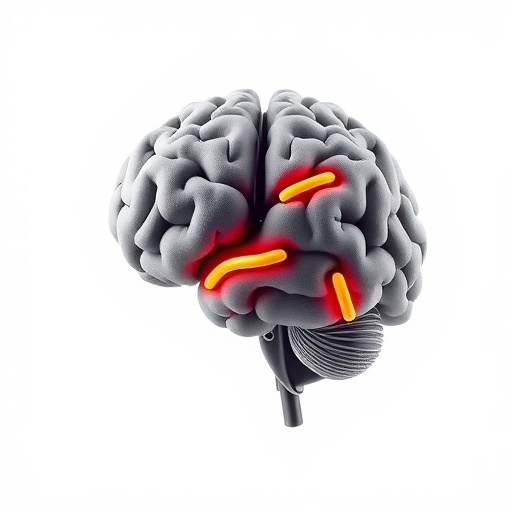
A groundbreaking study published recently in npj Parkinson’s Disease uncovers a compelling neurobiological link between REM sleep behavior disorder (RBD) and Parkinson’s disease (PD), marked by increased frontal binding of the radioligand [^123I]FP-CIT. This marker, traditionally used to assess presynaptic dopamine transporter (DAT) density, reveals unexpected patterns in PD patients who report RBD symptoms, offering fresh insights into disease heterogeneity and progression. As Parkinson’s disease continues to challenge neurologists and researchers alike with its complex symptomatology and unclear pathophysiology, this novel finding could reshape how clinicians understand and potentially monitor the non-motor manifestations of PD.
Parkinson’s disease is widely known for its hallmark motor symptoms—tremor, bradykinesia, rigidity—rooted in degeneration of dopaminergic neurons in the nigrostriatal pathway. However, non-motor symptoms such as sleep disturbances, particularly REM sleep behavior disorder, frequently precede motor deficits by years, complicating clinical assessments. RBD, characterized by loss of normal muscle atonia during REM sleep, causes individuals to physically enact their dreams, often violently. This behavior is more than a nocturnal oddity; it’s increasingly recognized as a prodromal marker for synucleinopathies like PD. Understanding neural correlates of RBD in PD could illuminate prodromal or early disease mechanisms and pave the way for earlier interventions.
The current study employed [^123I]FP-CIT Single Photon Emission Computed Tomography (SPECT), a neuroimaging modality widely implemented to quantify dopamine transporter availability, which reflects dopaminergic nerve terminal integrity. While prior studies have extensively documented reduced striatal DAT binding in PD reflecting dopaminergic neurodegeneration, this investigation focused on extrastriatal binding patterns, particularly in the frontal cortex. The frontal cortex, integral to executive function, mood regulation, and behavioral control, has rarely been implicated directly in dopaminergic imaging assessments of PD. Thus, increased frontal binding in patients presenting with self-reported RBD suggests previously uncharted neurochemical dynamics.
Methodologically, the cohort comprised Parkinson’s disease patients who self-identified their RBD status rather than being formally diagnosed through polysomnography. Despite this limitation, the self-reports correlated strongly with distinct imaging signatures. The analysis revealed that those reporting RBD symptoms exhibited significantly increased frontal [^123I]FP-CIT binding relative to PD patients without such reports and to healthy controls. This contrast underscores a possible compensatory or pathological upregulation of dopamine transporter expression or availability in frontal regions, which might be intimately connected with RBD pathophysiology in PD.
This novel observation confounds the traditional notion of uniform dopaminergic loss across PD-affected brain regions. Instead, it hints at a regional heterogeneity where frontal cortical areas could retain or augment tonic dopaminergic function in association with REM sleep disturbances. The implications are profound: frontal DAT density might serve as a biomarker for phenotypic subtypes of PD, where RBD symptoms demarcate distinct neurochemical trajectories or compensatory mechanisms within the dopaminergic system.
Dopamine’s role in sleep regulation, especially REM sleep, is complex and multifaceted. Preclinical studies have long suggested dopamine modulates REM sleep through subcortical and cortical circuits, influencing neuronal excitability and arousal thresholds. In the context of PD, where dopaminergic loss is pronounced and multifocal, alterations in dopamine dynamics in the frontal cortex may disrupt normal REM atonia mechanisms, facilitating the emergence of RBD. Elevated [^123I]FP-CIT binding detected in this study may reflect an adaptive response to dopaminergic neuronal loss elsewhere or represent aberrant dopaminergic signaling underlying abnormal REM sleep behavior.
Furthermore, the study’s insights align intriguingly with emerging concepts of synucleinopathy progression. Lewy body pathology, the principal driver in PD, exhibits a caudo-rostral spread from lower brainstem structures implicated in REM sleep regulation to cortical regions. Increased frontal DAT binding may represent early or compensatory synaptic changes consequent to this progression, correlating with clinical RBD manifestation. If this link is consistently replicated, imaging the frontal cortex could become a critical component for staging PD and stratifying patients based on RBD presence or absence.
Delving into the dopaminergic pharmacology underpinning these findings, [^123I]FP-CIT binds with high affinity to dopamine transporters but also interacts, albeit less selectively, with serotonin transporters. Hence, increased frontal binding might also involve serotonergic contributions or upregulation, which require further investigation. This interplay between dopaminergic and serotonergic systems could be pivotal in the neurobiology of RBD and sleep disturbances in Parkinson’s disease, illuminating new therapeutic targets beyond dopamine replacement therapies.
Clinically, the identification of increased frontal [^123I]FP-CIT binding in PD patients with RBD holds promise for refining diagnostic accuracy. Since polysomnography is resource-intensive and not universally accessible, imaging biomarkers that correlate robustly with RBD symptoms could expedite phenotyping and risk stratification. This is especially valuable for predicting prognosis as RBD presence associates with more rapid motor deterioration, cognitive decline, and other non-motor symptoms in PD, portending a more aggressive disease course.
Moreover, the study opens avenues for longitudinal research to ascertain whether frontal DAT alterations precede clinical RBD onset or follow its manifestation. Understanding temporal dynamics could provide critical windows for intervention and pave the way for preventative or disease-modifying strategies targeted to dopaminergic circuits. Imaging biomarkers such as [^123I]FP-CIT SPECT might eventually integrate with genetic, clinical, and neurophysiological data to generate comprehensive predictive models for PD progression.
The methodology utilized also emphasizes the utility of advanced neuroimaging in neurodegenerative disease research. By extending analyses beyond traditional striatal regions and embracing whole-brain assessments, researchers can uncover subtler, region-specific neurochemical alterations that underlie diverse symptom clusters. This approach may catalyze a paradigm shift away from monolithic views of PD towards recognizing its multifaceted neurobiological heterogeneity.
Despite its provocative findings, the study underscores the necessity for cautious interpretation. Self-reported RBD, while practical, lacks the diagnostic rigor of polysomnographic confirmation. Future studies incorporating objective sleep measures and larger cohorts are essential to validate and extend these findings. Additionally, disentangling the contributions of dopamine versus serotonin transporter binding in the frontal cortex will require multimodal imaging and pharmacological challenge studies.
As neuroscientists and clinicians work toward unraveling the intricacies of Parkinson’s disease, this research injects fresh vitality into an area demanding urgent clarity. REM sleep behavior disorder is not merely a troublesome nocturnal phenomenon but a window into the brain’s evolving pathology. The discovery of augmented frontal [^123I]FP-CIT binding in these patients heralds new possibilities for understanding and ultimately mitigating the impact of PD’s non-motor symptoms that profoundly affect patient quality of life.
In summary, this study pioneers the detection of increased frontal dopamine transporter availability in Parkinson’s patients manifesting RBD, challenging conventional views of dopaminergic loss and regional pathology. The findings highlight the intricate relationship between sleep disorders and neurodegeneration, emphasizing the frontal cortex’s underappreciated role in PD. Such insights are crucial for developing nuanced biomarkers and personalized interventions, moving the field closer to precision medicine in Parkinson’s disease.
The road ahead invites expansive research to integrate these imaging biomarkers with molecular, genetic, and clinical data sets, forging a multidimensional understanding of PD subtypes. Ultimately, this may revolutionize early diagnosis, monitoring, and therapeutic response assessment, transforming lives affected by Parkinson’s disease.
Subject of Research: Parkinson’s disease, REM sleep behavior disorder, dopamine transporter imaging, neurodegeneration, frontal cortex neurochemistry.
Article Title: Increased frontal [^123I]FP-CIT binding in Parkinson’s disease patients with self-reported REM sleep behavior disorder.
Article References:
Jaakkola, E., Ijas, J., Joutsa, J. et al. Increased frontal [^123I]FP-CIT binding in Parkinson’s disease patients with self-reported REM sleep behavior disorder. npj Parkinsons Dis. 11, 243 (2025). https://doi.org/10.1038/s41531-025-01116-7
Image Credits: AI Generated


