In recent years, coronary artery disease (CAD) has emerged as a growing global health concern, with its incidence rising significantly across diverse populations. This widespread increase has prompted researchers to carefully examine the diagnostic tools used in identifying and managing CAD, particularly focusing on the safety and efficacy of these approaches. Among the most revealing findings from a recent comprehensive study is the pronounced variability in radiation exposure patients receive during diagnostic imaging procedures for CAD, a factor that has profound implications for patient health worldwide.
Computed tomography angiography (CTA), a pivotal imaging modality in diagnosing CAD, employs ionizing radiation to generate detailed images of coronary arteries. Despite its clinical advantages, CTA’s reliance on radiation presents inherent risks, particularly cumulative exposure that can predispose patients to adverse effects, including the potential development of radiation-induced diseases. The degree of radiation delivered during CTA, however, fluctuates considerably depending on technical parameters, operator expertise, and institutional protocols, leading to a lack of standardization that compromises patient safety on a global scale.
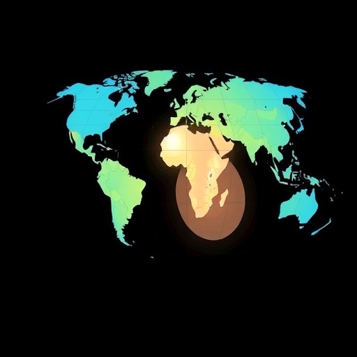
This variability is especially pronounced in low- and middle-income countries, where resource constraints often limit access to state-of-the-art imaging equipment and specialized training for medical personnel. In such settings, outdated machinery and suboptimal procedural protocols contribute to higher radiation doses during imaging, exacerbating the risk to vulnerable patient populations. Consequently, disparities in diagnostic radiation exposure underscore the urgent need to address inequalities in healthcare infrastructure and training globally.
At the heart of these discrepancies lies a technological landscape in flux. Advances in scanner hardware and software algorithms have enabled dose reduction strategies in high-income countries, yet these innovations remain underutilized in many regions. Iterative reconstruction techniques, optimized scanning protocols, and dose modulation features have been shown to significantly minimize radiation burden without compromising image quality. Their inconsistent application worldwide continues to fuel unnecessary radiation exposure during CAD diagnosis.
Moreover, operator-related factors accentuate the radiation dose variability. Radiologists and technologists wield substantial influence over procedural parameters, including scan range, tube current, and image acquisition phases. Lack of comprehensive training or standardized protocols may result in overexposure, particularly when the clinical imperative to obtain diagnostic clarity overshadows radiation safety principles. Bridging this expertise gap is essential to harmonizing radiation doses across diverse healthcare environments.
The study’s findings also raise critical ethical considerations regarding informed consent and patient communication. Patients undergoing coronary CTA must be apprised of the benefits and potential risks associated with radiological examination, including radiation dose implications. Transparent discussions are vital to empower patients to participate actively in decision-making about their diagnostic care, especially when alternative non-ionizing imaging options exist or screening appropriateness is in question.
From a policy perspective, international collaboration is indispensable in establishing global standards for radiation use in cardiac imaging. Regulatory frameworks tailored to harmonize dose thresholds and quality assurance measures can drive improvements in equipment procurement, operator training, and procedural implementation. Such initiatives would promote equitable healthcare delivery and mitigate preventable radiation risks, aligning with broader public health objectives.
In addition, ongoing research into novel diagnostic modalities holds promise for reducing reliance on ionizing radiation. Magnetic resonance angiography (MRA) and advanced ultrasound technologies offer radiation-free alternatives with evolving diagnostic capabilities. Integrating these approaches into clinical pathways, especially for screening and follow-up evaluations, may help attenuate the cumulative radiation burden on CAD patients worldwide.
The study also emphasizes the critical role of continuous education and quality improvement initiatives. Medical institutions must prioritize regular training programs focusing on radiation safety, protocol optimization, and dose monitoring. Implementation of audit systems and dose registries can foster accountability and facilitate benchmarking, accelerating the adoption of best practices that safeguard patient health.
Furthermore, advancements in artificial intelligence (AI) technologies present an emerging frontier in dose optimization. AI-driven algorithms can assist operators in tailoring scan parameters dynamically, enhancing image quality while minimizing radiation exposure. Deployment of such intelligent systems, particularly in resource-limited settings, could revolutionize CAD diagnostics by compensating for gaps in human expertise and equipment limitations.
Addressing these multifaceted challenges requires a coordinated approach encompassing stakeholders from clinical medicine, medical physics, healthcare policy, and technology development. Investment in infrastructure upgrades, capacity building, and international knowledge exchange are fundamental components driving sustainable improvement in diagnostic radiation use. Without concerted efforts, the global disparity in radiation exposure risks exacerbating health inequities related to CAD outcomes.
In conclusion, the marked variation in radiation doses administered during coronary diagnostic testing illuminates a critical area for intervention in modern cardiology. Reducing unnecessary radiation exposure through updated equipment, standardized protocols, and enhanced operator training is imperative—especially in economically disadvantaged regions. By leveraging technological innovations and fostering global collaboration, the medical community can significantly improve the quality and safety of CAD diagnosis, ultimately contributing to better patient outcomes on a worldwide scale.
Subject of Research: Coronary artery disease diagnostic imaging and radiation dose variability
Article Title: Not available
News Publication Date: Not available
Keywords
Radiation, Coronary artery disease, Diagnostic imaging, World population, Tomography, Angiography, Medical equipment
