In a groundbreaking advancement that could reshape the landscape of Parkinson’s disease research, a recent study delves into a novel biomarker offering unprecedented insight into mild cognitive impairment (MCI) associated with this neurodegenerative condition. Researchers Chen, Liu, Kou, and their colleagues have identified free water levels in the external globus pallidus as a compelling predictor of MCI in Parkinson’s patients. Published in the esteemed journal npj Parkinson’s Disease, their findings not only illuminate the underlying neurological transformations but also establish a critical connection to serum neurofilament light chain (NfL) levels, a protein indicative of neuronal damage.
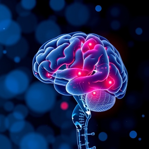
Parkinson’s disease, predominantly known for its motor symptoms such as tremors and rigidity, carries a significant burden of non-motor complications including cognitive decline, which profoundly affects patient quality of life. Early detection of cognitive impairment remains an urgent challenge, as current diagnostic paradigms often miss subtle changes before irreversible damage occurs. The external globus pallidus (GPe), a component deep within the brain’s basal ganglia complex, has long been implicated in the modulation of movement and cognitive functions. However, this study brings to light its potential as a biomarker reservoir through the quantification of extracellular free water.
Advanced neuroimaging techniques, particularly diffusion magnetic resonance imaging (dMRI), were pivotal in quantifying the free water fraction within the GPe. This free water measure reflects extracellular fluid alterations, which can signify neuroinflammation, edema, or neuronal loss. Elevated free water levels in the GPe emerged as a potent harbinger of developing MCI, marking neural tissue microenvironment changes that precede overt clinical symptoms. This biomarker thus provides a window into the pathophysiological processes shaping cognitive decline in Parkinson’s disease.
What sets this research apart is the dual focus on both neuroimaging and peripheral biomarkers. Serum neurofilament light chain, a cytoskeletal protein released into the bloodstream following axonal injury, offers a minimally invasive proxy for neurodegeneration. The researchers discovered a robust association between increased GPe free water and elevated serum NfL, suggesting that extracellular fluid changes in the basal ganglia mirror systemic neuronal damage detectable in blood samples. This correlation paves the way for integrating brain imaging with blood-based assays in comprehensive Parkinson’s disease monitoring.
The implications of these findings extend well beyond diagnostic enhancement. Understanding free water alterations in the GPe could unveil new therapeutic targets aimed at mitigating or delaying cognitive deterioration. Neuroinflammation, a likely contributor to increased free water, represents a modifiable pathophysiological axis. Agents designed to reduce neuroinflammatory processes or stabilize extracellular fluid homeostasis might preserve cognitive function if administered in early disease phases.
Moreover, this research adds a nuanced layer to the complex interplay of neural circuits impacted by Parkinson’s. The basal ganglia, traditionally studied for their role in movement, are increasingly recognized for cognitive integration. Disruptions in the GPe’s microenvironment, evidenced by elevated free water, could perturb the delicate balance of excitatory and inhibitory signaling crucial for cognitive processing. This insight enhances mechanistic models of Parkinson’s related cognitive decline, refining targets for future interventional studies.
Critically, the application of free water imaging circumvents limitations intrinsic to other biomarkers fraught with variability or invasiveness. Unlike conventional MRI markers, which primarily reflect structural atrophy, free water measures capture subtle extracellular changes that precede anatomical loss. Concurrently, serum NfL levels provide accessible, repeatable measures, enabling longitudinal tracking of disease progression and treatment response. The convergence of these modalities exemplifies precision medicine approaches tailored to individual patient trajectories.
The study’s methodology involved a considerable cohort of Parkinson’s patients stratified by cognitive status. Through rigorous statistical analyses controlling for demographic and clinical variables, the association between GPe free water and MCI remained highly significant. The reproducibility of these findings across independent samples further affirm their robustness, underscoring the biomarker’s potential for clinical utility. Researchers advocate for larger, multicenter trials to validate and standardize free water quantification protocols.
From a technological perspective, advancements in diffusion imaging sequences and analytical algorithms were crucial for the sensitive detection of free water variations. These innovations minimize confounds such as partial volume effects and motion artifacts, enhancing the fidelity of measurement. As imaging platforms continue to evolve, accessibility to high-resolution diffusion data is becoming increasingly feasible in clinical settings, accelerating translational adoption.
Beyond the immediate context of Parkinson’s disease, this research invites exploration of free water dynamics in other neurodegenerative disorders characterized by cognitive decline, such as Alzheimer’s disease and multiple system atrophy. Comparative studies may reveal disease-specific patterns of extracellular fluid disturbances, broadening the biomarker’s applicability and enriching our understanding of neurodegeneration’s diverse pathological landscapes.
Ethical considerations accompany the promise of early detection biomarkers. Identifying patients at risk for cognitive impairment before symptoms manifest raises questions about patient counseling, psychological impact, and therapeutic options. However, a proactive approach grounded in scientifically validated biomarkers empowers clinicians and patients, facilitating timely interventions and potentially altering disease trajectories.
In conclusion, the elucidation of free water content in the external globus pallidus as a predictor of mild cognitive impairment in Parkinson’s disease marks a seminal advance in neurodegenerative research. This marker’s interplay with serum neurofilament light chain levels bridges central and peripheral manifestations of neuronal injury, offering a multifaceted perspective on disease mechanisms. As this research matures, it promises to refine diagnostic accuracy, inform therapeutic development, and ultimately improve outcomes for millions grappling with Parkinson’s disease worldwide.
Subject of Research: Biomarkers predicting mild cognitive impairment in Parkinson’s disease, focusing on free water levels in the external globus pallidus and their relationship with serum neurofilament light chain.
Article Title: Free water in the external globus pallidus predicts mild cognitive impairment in Parkinson’s disease and is associated with serum neurofilament light chain levels.
Article References:
Chen, H., Liu, H., Kou, W. et al. Free water in the external globus pallidus predicts mild cognitive impairment in Parkinson’s disease and is associated with serum neurofilament light chain levels. npj Parkinsons Dis. (2026). https://doi.org/10.1038/s41531-026-01291-1
Image Credits: AI Generated
