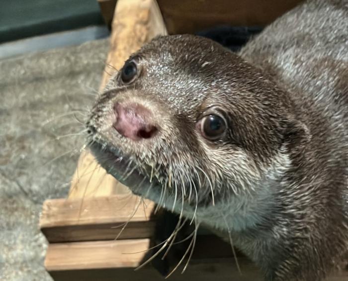
In a remarkable case reported by Osaka Metropolitan University, an 11-year-old neutered male Asian small-clawed otter, a species known scientifically as Aonyx cinereus, fell down the stairs while sleeping, leading to an unexpected and severe medical condition: left-sided paralysis. This incident has raised important discussions about animal care, rehabilitation, and the potential underlying causes of acute neurological symptoms in wildlife.
Upon the onset of paralysis, the otter underwent an initial treatment regimen involving prednisolone, carefully administered at a dose of 0.5 mg/kg once daily. Prednisolone, a synthetic corticosteroid, is commonly used in veterinary medicine to reduce inflammation and suppress the immune response. The otter’s caregivers hoped that this treatment would alleviate the symptoms and promote recovery, illustrating the delicate balance of providing immediate aid while seeking a comprehensive diagnosis.
Despite the otter displaying minor improvements by day 10 of the treatment, the paralysis continued unabated. This stagnation prompted the veterinary team to utilize advanced imaging techniques for a deeper investigation into the otter’s condition. Magnetic Resonance Imaging (MRI) was performed, specifically focusing on T2-weighted imaging (T2WI), which proved critical in diagnosing the issue. The results unveiled a well-defined, hyperintense lesion located on the left side of the spinal cord, specifically at the C2-3 intervertebral level.
The presence of this lesion raised the suspicion of Fibrocartilaginous Embolus (FCE), a serious condition where small fragments of cartilage can obstruct blood flow to the spinal cord, leading to ischemic injury. This diagnosis is particularly important as FCE can often present without the expected signs of deterioration, making it challenging for veterinarians to identify the condition promptly.
With the diagnosis in hand, the veterinary team adjusted the treatment protocol by tapering the prednisolone, a decision rooted in the understanding of corticosteroids’ role in managing inflammatory responses while allowing the body to recover naturally. Remarkably, by day 23, the otter was able to walk normally, a significant milestone that warranted the discontinuation of corticosteroid administration. This positive turnaround served as a testament to the resilience of the species and the effectiveness of prompt and appropriate veterinary intervention.
Interestingly, the progress did not halt there. A follow-up MRI conducted one year later demonstrated a significant reduction in the size of the lesion compared to the initial examination, hinting at successful healing processes and the possibility of functional recovery in the otter’s neurological pathways. This long-term follow-up highlights not only the potential benefits of acute identification and treatment but also the importance of ongoing monitoring to assess the full scope of recovery.
These findings underscore a pivotal point in veterinary medicine, particularly in the caring for exotic and wild species: when faced with acute paralysis and neurologic deficits in an animal, the consideration of fibrocartilaginous embolus should be paramount. Such insights could lead to improved diagnostic patterns and treatment protocols, enriching the current body of knowledge required for effective wildlife rehabilitation.
The case emphasizes a nexus between veterinary science, wildlife conservation, and animal welfare. By dissecting complex medical issues and sharing these findings, researchers and veterinarians alike contribute to a more profound understanding of animal health, promoting better outcomes for countless creatures who may find themselves in similar predicaments.
As researchers continue to evaluate similar cases through rigorous scientific methods, the information gleaned from such instances, including this unforgettable experience involving the Asian small-clawed otter, enriches our knowledge of animal biology and affirms the necessity of specialized care. Such dedication not only benefits individual animals but also enhances the strategies employed across veterinary practices to ensure the well-being of diverse species in their care.
The broader implications extend beyond individual animal cases; they provoke a reevaluation of how veterinary science intersects with public awareness of wildlife health. An informed community that understands these issues is crucial to effective conservation efforts. With furor around animal welfare growing, public engagement through educational outreach becomes essential in mobilizing support for wildlife and their habitats.
This incident serves as a reminder of the unpredictable and delicate nature of wildlife care, emphasizing the critical role of veterinary expertise in identifying and responding to acute health crises effectively. Continuous advancements in medical imaging, diagnostics, and treatment options herald a promising future for wildlife rehabilitation.
By sharing these compelling narratives of recovery and resilience, there may emerge a stronger collective commitment toward preserving the health of wildlife populations, leading ultimately to richer biodiversity and a well-informed public better equipped to appreciate the wonders of nature.
Subject of Research: Fibrocartilaginous embolus in Asian small-clawed otters
Article Title: Suspected fibrocartilaginous embolus in Asian small-clawed otter (Aonyx cinereus)
News Publication Date: 7-Feb-2025
Web References: Osaka Metropolitan University
References: Journal of Veterinary Medical Science
Image Credits: Osaka Metropolitan University
Keywords: Asian small-clawed otter, left-sided paralysis, fibrocartilaginous embolus, veterinary medicine, MRI, rehabilitation, wildlife care, animal health, neurological symptoms.


