In a groundbreaking study poised to reshape our understanding of neurodegenerative diseases, researchers have uncovered a compelling inverse relationship between limbic alpha-synucleinopathy and myelin integrity in both murine models and human subjects. Published in the forefront journal npj Parkinson’s Disease, this investigation spearheaded by Clark, Landes, Abbas, and colleagues elaborates on the complex interplay between pathological alpha-synuclein accumulation and the white matter architecture critical for neural function.
Parkinson’s disease, alongside related synucleinopathies, is characterized by the aberrant deposition of alpha-synuclein protein aggregates within the nervous system. Traditionally, the research community has focused predominantly on neuronal loss and dopamine deficiency. However, this novel study shifts the spotlight towards the myelin sheath—an insulating layer surrounding axons made by oligodendrocytes—implicating its perturbation in disease progression and symptomatology with unprecedented granularity.
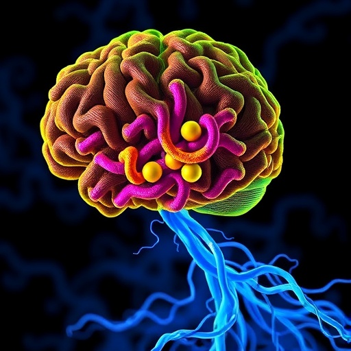
The limbic system, encompassing vital brain regions such as the hippocampus and amygdala, is essential for emotional processing, memory formation, and autonomic regulation. Prior findings have implicated limbic alpha-synuclein pathology in both the prodromal and symptomatic phases of Parkinson’s disease and dementia with Lewy bodies. Yet, little was understood about how alpha-synuclein accumulation interacts with the structural and functional state of myelin, until now.
Clark and colleagues employed a comprehensive battery of molecular, histological, and imaging techniques across genetically engineered mouse models exhibiting limbic alpha-synucleinopathy, complemented by postmortem analysis of human brain tissues from Parkinson’s patients. Their multi-modal approach allowed for the quantification of myelin proteins, such as myelin basic protein (MBP) and proteolipid protein (PLP), within regions exhibiting high alpha-synuclein burden.
Intriguingly, the results revealed a robust inverse correlation: areas with pronounced alpha-synuclein aggregation demonstrated markedly reduced expression of key myelin markers. This reduction was not merely a coincident observation but appeared mechanistically linked, suggesting that pathological alpha-synuclein species may disrupt oligodendrocyte function or myelin maintenance. The data hint at a cascading effect where synucleinopathy exacerbates white matter damage, potentially impairing neural conductivity and synaptic efficiency critical for limbic network dynamics.
Further molecular analyses pinpointed altered signaling pathways within oligodendrocytes under stress from alpha-synuclein toxicity. Differential expression of genes involved in myelin synthesis and turnover was noted, alongside inflammatory mediators potentially contributing to a hostile local microenvironment. These changes likely compromise the regenerative capacity of myelin and amplify neurodegeneration.
The study also illuminated distinct temporal dynamics in which myelin deterioration follows or parallels alpha-synuclein pathology onset. Longitudinal assessments in the mouse models suggested a possible window for therapeutic intervention aimed at preserving myelin integrity before irreversible neuronal loss ensues. This challenges the traditional neuron-centric therapeutic approaches dominating current clinical strategies.
Human tissue analysis corroborated murine findings, underscoring translatability and clinical relevance. Notably, decreases in MBP and other myelin-associated proteins were observed prominently in limbic areas densely populated by Lewy bodies and Lewy neurites, hallmark structures consisting mainly of alpha-synuclein fibrils. These findings resonate with emerging hypotheses implicating white matter disruption in cognitive and psychiatric manifestations frequently observed in Parkinson’s disease beyond the classical motor symptoms.
From a clinical perspective, this research opens avenues for novel biomarkers leveraging myelin integrity as a surrogate for early disease diagnosis and progression monitoring. Advanced neuroimaging modalities sensitive to myelin changes, such as magnetization transfer imaging and myelin water fraction mapping, could become invaluable tools in future trials assessing disease-modifying agents.
Moreover, the insights provoke reconsideration of therapeutic strategies to encompass not only alpha-synuclein clearance or aggregation inhibition but also interventions promoting oligodendrocyte survival, myelin repair, and neuroinflammation modulation. Such a multifaceted approach may enhance functional outcomes and slow the relentless clinical decline afflicting patients.
Mechanistically, the interaction between alpha-synuclein and myelin biology might involve direct aggregation effects on oligodendrocytes or indirect consequences mediated by microglia and astrocytes. The study discusses plausible pathways including proteostasis disruption, mitochondrial dysfunction, and oxidative stress, all converging to weaken the white matter scaffold underpinning neuronal connectivity.
The research has profound implications for understanding the heterogeneity and spectrum of Parkinson’s disease presentations, where some patients exhibit disproportionately severe cognitive and limbic impairments. This limbic alpha-synucleinopathy-associated myelin loss could underlie these phenotypic variations, offering new targets for personalized medicine approaches.
Future research directions, as outlined by Clark et al., prioritize dissecting the molecular mechanisms linking alpha-synuclein accumulation to oligodendrocyte pathology, including the role of extracellular vesicles and prion-like propagation of misfolded protein aggregates. Investigating the potential reversibility of myelin damage also emerges as a critical step towards designing reparative therapies.
In sum, this seminal study furnishes compelling evidence positioning myelin disruption as a pivotal and perhaps underestimated facet of limbic alpha-synucleinopathies. By bridging murine genetic models with human neuropathological validation, the investigators chart an innovative path forward in unraveling the complex cellular choreography underlying Parkinson’s disease and related disorders.
The paradigm shift heralded by Clark and colleagues mandates a holistic reassessment of neurodegeneration models, integrating neuronal and glial elements in a concerted landscape of disease pathology. Such integrative perspectives could expedite the development of combinatory treatments targeting multifactorial mechanisms driving clinical decline, thereby improving patient outcomes in synucleinopathies and beyond.
As this field evolves, the identification of reliable myelin-related biomarkers alongside alpha-synuclein metrics will revolutionize early diagnosis and therapeutic tracking. The insights also underscore the need for interdisciplinary collaboration, leveraging advances in molecular neuroscience, neuroimaging, and clinical neurology to translate these findings from bench to bedside.
In conclusion, the elucidation of an inverse link between limbic alpha-synucleinopathy and myelin markers marks an epochal advancement. It underscores myelin’s centrality to neurodegenerative disease mechanisms and highlights novel targets for intervention that may ultimately alter the trajectory of Parkinson’s disease and cognate disorders, offering renewed hope to millions worldwide.
Subject of Research:
The study addresses the pathological relationship between alpha-synuclein accumulation in limbic brain regions and the concurrent disruption of myelin-associated proteins, exploring mechanisms underlying neurodegeneration in Parkinson’s disease and related synucleinopathies through combined animal and human analyses.
Article Title:
Testing an inverse link between limbic alpha-synucleinopathy and myelin markers in mice and humans.
Article References:
Clark, R.N., Landes, R.E., Abbas, M. et al. Testing an inverse link between limbic alpha-synucleinopathy and myelin markers in mice and humans. npj Parkinsons Dis. (2026). https://doi.org/10.1038/s41531-026-01278-y
Image Credits:
AI Generated
