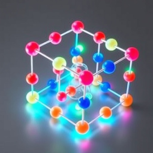
In a groundbreaking advance in the field of photonics and bioimaging, researchers have developed an innovative method to engineer ultrasound-responsive phosphorescence in aqueous solutions by constructing microscale rigid frameworks around carbon nanodots (CNDs). This pioneering work, led by Liang Yachuan and colleagues from Zhengzhou University of Light Industry in collaboration with Liu Kaikai’s team from Zhengzhou University, unveils a sophisticated strategy that enhances the stability and activation of triplet excitons—long-lived excited states critical for durable phosphorescence—by harnessing the self-assembly properties of cyclodextrin under ultrasonic stimulation. Their findings, recently published in the esteemed journal Light: Science & Applications, chart new territory in the development of stimuli-responsive room temperature phosphorescent (RTP) materials with promising applications in ultrasound detection and in vivo imaging.
Triplet excitons, characterized by their extended lifetimes, are fundamental to the exceptional luminescent behavior observed in RTP materials. Unlike conventional fluorescence, which often lasts mere nanoseconds, the longevity of triplet excitons enables photons to be emitted over longer periods, significantly amplifying tissue imaging signal-to-noise ratios and facilitating deeper tissue penetration. These attributes make RTP materials especially valuable for non-invasive biomedical applications, where clear and enduring optical signals are paramount. However, the effective utilization of triplet excitons has been historically hindered by their susceptibility to rapid non-radiative dissipation caused by molecular vibrations, environmental quenchers like oxygen and water, and inherently weak spin–orbit coupling in many organic systems.
Addressing these challenges, the researchers introduced a micro-scale rigid framework engineering approach that fundamentally suppresses the non-radiative pathways typically responsible for triplet exciton loss. Their technique leverages the ultrasonic-initiated self-assembly of cyclodextrins, cyclic oligosaccharides known for their unique host-guest chemistry and propensity to form ordered supramolecular structures in aqueous media. Under ultrasonic agitation, cyclodextrins organize into crystalline frameworks that tightly encapsulate carbon nanodots, effectively immobilizing them within a rigid network. This confinement restricts molecular motions and environmental access that would otherwise quench triplet states, thereby dramatically enhancing RTP performance.
The innovation lies not only in the enhanced RTP efficiency but also in the material’s remarkable sensitivity to ultrasound stimuli, reflected in a prolonged phosphorescence lifetime of up to 1.25 seconds within aqueous environments. This ultrasound responsiveness is a direct consequence of the dynamic structural modulation of the cyclodextrin framework under varying ultrasonic intensities. As the crystalline rigidity of the cyclodextrin matrices increases, so does the degree of triplet exciton stabilization and the observable phosphorescent emission. This controllable responsiveness paves the way for the development of sophisticated optical sensors and imaging agents capable of real-time environmental feedback.
The researchers further explored the tunability of these RTP carbon dots by exploiting Förster resonance energy transfer (FRET) mechanisms within their system. FRET facilitated the realization of multi-color afterglow emissions modulated by ultrasonic inputs in aqueous solutions, thereby expanding the versatility and potential usability of these materials in diverse analytical and diagnostic applications. The ability to fine-tune emission colors dynamically under ultrasound stimulation introduces unprecedented opportunities for multiplexed imaging and advanced data encryption technologies based on time-resolved afterglow signatures.
Importantly, the ultrasound-responsive phosphorescent carbon dots display exceptional promise for biomedical applications, such as ultrasound radar detection and in vivo afterglow imaging. Their prolonged lifetimes and high sensitivity to external acoustic stimuli allow for enhanced contrast imaging with minimized background noise interference—a critical advantage over conventional fluorescent probes that suffer from rapid decay and interference from tissue autofluorescence. This research thus represents a significant stride toward the practical deployment of RTP materials in clinical and environmental monitoring contexts.
The work also highlights key technical challenges intrinsic to the development of stimuli-responsive RTP materials. Historically, the rapid deactivation of triplet excitons through non-radiative relaxation and quenching by molecular oxygen has posed formidable barriers. The dual requirement of simultaneously regulating both the triplet excitons and the responsive stimulus sites further compounds material design complexity. By integrating the self-assembly dynamics of cyclodextrin with ultrasonic modulation, the researchers surmounted these obstacles, realizing a tunable system that balances exciton longevity with external sensitivity.
Fundamentally, the use of cyclodextrin as a supramolecular scaffold presents a compelling material paradigm due to its abundant hydrogen bonding capacity and structural versatility. Incorporating carbon nanodots within these host frameworks restricts intramolecular vibrations and shields excitons from external quenchers—a principle that may translate to broader classes of luminescent materials beyond carbon-based systems. The method’s scalability and compatibility with aqueous media mark significant steps toward biocompatible, environmentally benign RTP technologies.
The remarkable 1.25-second phosphorescence lifetime achieved in aqueous solution underscores the efficacy of rigid framework engineering at the microscale, a method that contrasts with prior approaches relying on covalent or multiple non-covalent interactions that often complicate synthesis and limit responsiveness. This research effectively demonstrates that mechanical agitation via ultrasound—an easily accessible, non-invasive external input—can be harnessed to dynamically regulate luminescent properties in real time, opening novel avenues for externally controllable optical devices.
Moreover, the team’s findings extend the frontiers of optical sensing by showcasing the potential of RTP materials to detect ultrasound signals with high sensitivity, enabling enhanced ultrasonic radar systems. Such applications may innovate remote sensing technologies across medical diagnostics, environmental monitoring, and industrial safety by leveraging the unique photophysical properties of these engineered nanomaterials.
In summary, this study delivers a transformative approach to engineering ultrasound-responsive RTP carbon nanodots via micro-scale rigid framework formation, overcoming longstanding limitations in triplet exciton stability and stimulus control. The integration of supramolecular chemistry, nanomaterial design, and acoustic modulation coalesces into a platform with far-reaching implications—ushering in a new generation of advanced luminescent materials poised to revolutionize bioimaging, sensing, and optoelectronics.
Subject of Research: Ultrasound-responsive room temperature phosphorescent carbon nanodots engineered via microscale rigid framework assembly
Article Title: Ultrasound-responsive phosphorescence in aqueous solution enabled by microscale rigid framework engineering of carbon nanodots
Web References:
DOI: 10.1038/s41377-025-01965-0
Image Credits: Yachuan Liang et al.
Keywords
Room temperature phosphorescence, triplet excitons, carbon nanodots, cyclodextrin self-assembly, ultrasound-responsive materials, microscale rigid frameworks, aqueous phosphorescence, non-radiative transition suppression, Förster resonance energy transfer, ultrasound imaging, bioimaging, optical sensing.


