In a groundbreaking advancement at the intersection of forensic science and artificial intelligence, researchers have unveiled an innovative deep learning approach designed to automate the detection of rib fractures on postmortem computed tomography (CT) scans. This novel methodology promises to revolutionize the field of forensic imaging by enhancing diagnostic precision, expediting medico-legal investigations, and ushering in a new era of computational pathology. Traditionally, the forensic examination of skeletal trauma, especially rib fractures, has relied heavily on manual assessment by experts, which is time-consuming, prone to human error, and inconsistently reproducible. The integration of deep learning techniques addresses these challenges directly by providing an automated, consistent, and highly sensitive detection system.
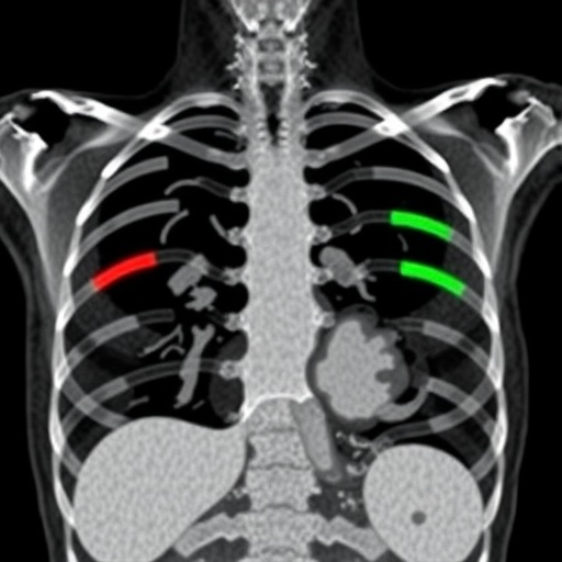
Rib fractures constitute a critical indicator in the forensic evaluation of trauma-related deaths, often shedding light on the circumstances surrounding fatal injuries. Postmortem CT imaging has increasingly become a valuable modality in forensic examinations due to its ability to non-invasively visualize the internal structure of the thoracic cage in exquisite detail. However, the sheer volume and complexity of CT data present substantial analytical burdens. This new study recognizes the urgent need for computational tools capable of navigating these challenges. By applying state-of-the-art deep learning algorithms, specifically designed convolutional neural networks (CNNs), the researchers have developed a robust framework that processes volumetric CT data to automatically identify rib fractures with high accuracy.
The design of the deep learning model involved training on a large dataset of postmortem CT scans, meticulously labeled by forensic radiologists to ensure ground-truth reference. This supervised learning paradigm enabled the network to discern intricate fracture patterns and subtle degenerative changes that often complicate manual evaluation. The model leverages advanced convolutional layers configured to capture three-dimensional contextual information, enhancing the system’s ability to differentiate between pathological rib deformities and genuine fractures caused by trauma. Early validation results demonstrate remarkable sensitivity and specificity metrics, evidencing the potential for clinical and forensic adoption.
One of the most compelling aspects of this research is the model’s applicability in real-world forensic workflows. The automation of rib fracture detection not only reduces the cognitive and operational workload of forensic experts but also minimizes inter-observer variability, a notable concern in forensic pathology. The researchers emphasize that their algorithm functions as an assistive tool designed to augment the decision-making process, thereby allowing forensic practitioners to focus on complex case interpretations and integrative analyses. Furthermore, the reduced time-to-diagnosis could expedite the entire death investigation process, providing timely information for legal scrutiny and family closure alike.
The integration of this AI-driven system holds significant implications beyond forensic medicine. Rib fractures are a common manifestation of blunt force trauma and can serve as crucial markers in clinical trauma care as well. Although the current application targets postmortem data, the underlying technology has the potential to be adapted for live clinical scenarios, offering immediate, automated fracture assessments in emergency and critical care settings. Such cross-domain applicability underscores the technological versatility and societal impact of this deep learning approach.
Technically, the implementation underscores several innovations in handling postmortem CT image data. The team tackled the challenge of heterogeneous image quality and variable artifact presence, which are typical in postmortem scans due to decomposition and handling conditions. Through sophisticated image pre-processing pipelines and augmentation strategies, the model’s input data was homogenized, allowing it to generalize across diverse cases. Importantly, the use of three-dimensional CNN architectures marks a significant leap from conventional two-dimensional slice-based analyses, facilitating a holistic interpretation of rib cage morphology and fracture connectivity.
The study also openly discusses the limitations and future directions. While the algorithm achieved outstanding performance in the training and validation sets, a multicentric study involving diverse forensic centers and wider demographic representation is necessary to confirm the generalizability of the results. Moreover, integrating the model with complementary forensic imaging modalities, such as magnetic resonance imaging or postmortem angiography, could enhance diagnostic fidelity by providing multimodal perspectives. The researchers express optimism that ongoing refinements and broader validation will cement AI’s role as an indispensable tool in forensic investigations.
Ethical considerations inherent to deploying AI in forensic contexts are thoughtfully addressed. The researchers highlight the importance of transparency and explainability, ensuring that forensic experts can scrutinize AI-generated outputs and maintain ultimate responsibility for case conclusions. They advocate for the establishment of standardized protocols governing the use of automated systems in medicolegal death investigations to ensure reproducibility and impartiality. This balanced approach aids in building professional and public trust in emerging forensic AI technologies.
Additionally, the development process showcased an inspiring synergy between domain experts and AI specialists. Extensive collaboration between forensic radiologists, pathologists, and machine learning engineers enabled a deep alignment of clinical relevance and technical prowess. Through iterative feedback loops, the model was tailored to address the nuanced anatomical and pathological features of rib fractures, all while maintaining computational efficiency. This multidisciplinary endeavor exemplifies the future trajectory of research initiatives aimed at harnessing AI’s full potential in medicine and law.
From a broader scientific standpoint, this study illuminates the transformative capacity of deep learning in analyzing complex biomedical images beyond traditional diagnostic tasks. The ability to detect fractures automatically from high-dimensional postmortem CT data ushers in a paradigm shift where machine intelligence complements human expertise. It champions a future where AI serves as a critical ally in extracting actionable insights from intricate anatomical data, potentially redefining standards of practice in forensic and clinical diagnostics.
Looking ahead, the researchers envision expanding the model’s scope to include a wider class of skeletal injuries, such as spinal fractures and cranial trauma. This generalized fracture detection toolkit could become an invaluable resource supporting forensic investigations comprehensively. Furthermore, the prospect of integrating natural language processing to generate preliminary forensic reports based on imaging findings is under exploration, which would significantly streamline case documentation and communication with judicial entities.
In sum, the automatic rib fracture detection system based on deep learning represents a monumental leap in forensic imaging technology. It combines cutting-edge artificial intelligence with detailed, large-scale postmortem CT data analysis to solve longstanding challenges in trauma diagnosis. As forensic medicine embraces digital transformation, innovations like this herald a future marked by increased accuracy, efficiency, and insight—ultimately enhancing the pursuit of justice and knowledge through science.
Subject of Research:
Article Title:
Article References:
Lopez-Melia, M., Magnin, V., Schranz, S. et al. Automatic rib fracture detection on postmortem CT data using deep learning. Int J Legal Med (2025). https://doi.org/10.1007/s00414-025-03669-x
Image Credits: AI Generated
DOI: https://doi.org/10.1007/s00414-025-03669-x
